Discover and read the best of Twitter Threads about #Radiology
Most recents (24)
Up for a Sunday tweetorial? 🤓
If you see this multicystic lung lesion 🫁 in the posterobasal region of the lower lobes in a young patient 🚹🚺with no pathological history, two differential diagnoses should be considered: CPAM vs pulmonary sequestration ✅
#radiology #FOAMrad
If you see this multicystic lung lesion 🫁 in the posterobasal region of the lower lobes in a young patient 🚹🚺with no pathological history, two differential diagnoses should be considered: CPAM vs pulmonary sequestration ✅
#radiology #FOAMrad

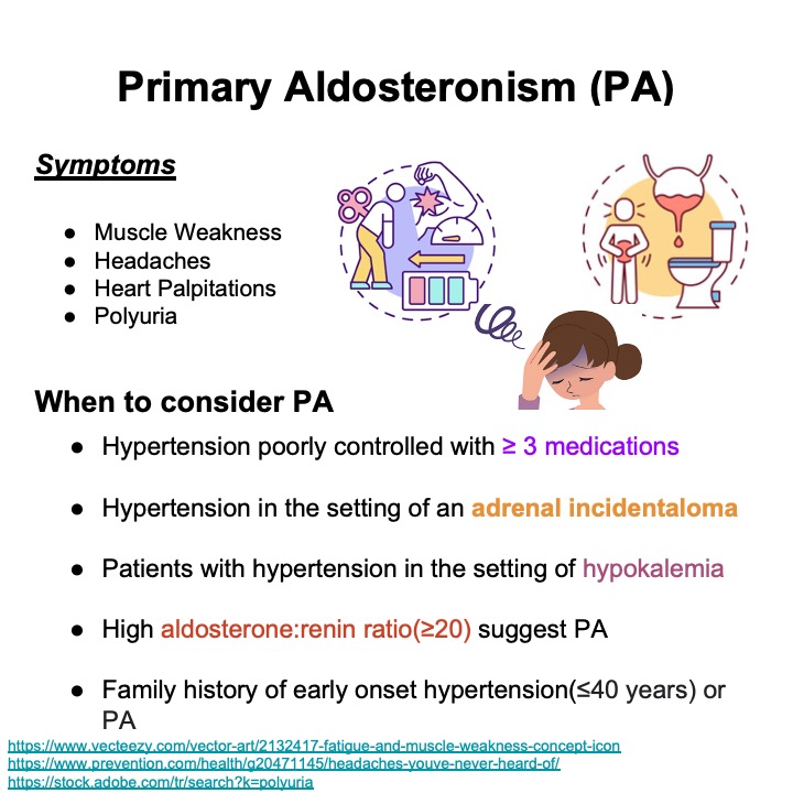
Check out this starter kit on Adrenal Vein Sampling created by MSC Reserves, Alperen Elek (@ElekAlperen), Lulu Zhang, MD, and Tina Chatterje ( @tinachatterje3)! #iRad #meded #radiology #interventionalradiology #radres #iradres 

Much has changed in my profession since I commenced family medicine practice in Saskatoon in 1972. Some of the changes that are now in progress or are pending are very positive. Others are disappointing. I'll focus on the positive changes first (1/18)
From the very outset of my career I was frustrated with Fee-For-Service (FFS) physician compensation as it tended to much more generously reward MDs focused primarily on procedural services & poorly compensate those spending time talking with patients (2/18)
In hope of addressing this compensation inequity, I became actively involved in leadership roles with the Saskatchewan Medical Association (SMA) culminating in my service as SMA President in 1979-80 (3/18)
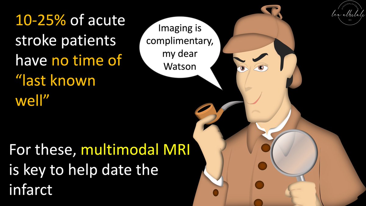
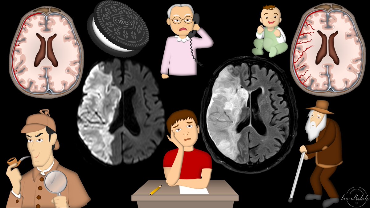
1/Time is brain! But what time is it?
If you don’t know the time of stroke onset, are you able to deduce it from imaging?
Here’s a #tweetorial to help you date a #stroke on MR!
#medtwitter #meded #neurotwitter #neurology #neurorad #radres #radtwitter #radiology #FOAMed #FOAMrad
If you don’t know the time of stroke onset, are you able to deduce it from imaging?
Here’s a #tweetorial to help you date a #stroke on MR!
#medtwitter #meded #neurotwitter #neurology #neurorad #radres #radtwitter #radiology #FOAMed #FOAMrad
A snapshot in time confounded by @TheNRMP match “algorithm”. The truth is #medicine is we have work to do in #genderequity - US physician workforce only 37% women. Rads 27%, Gen Surg 22%, #EM 👋🏼29%…@acepnow @WomenSurgeons @AmCollSurgeons @radiologyacr @EmergencyDocs @aawr_org… twitter.com/i/web/status/1…
@AAMCtoday stats on 2021 US physicians by % female. #pediatrics 65%, #orthopedics 5.9%. Note surgical sub speciality ($$$) higher male dominance, medical/primary care higher female…#orthotwitter @aap_peds @aaos1 
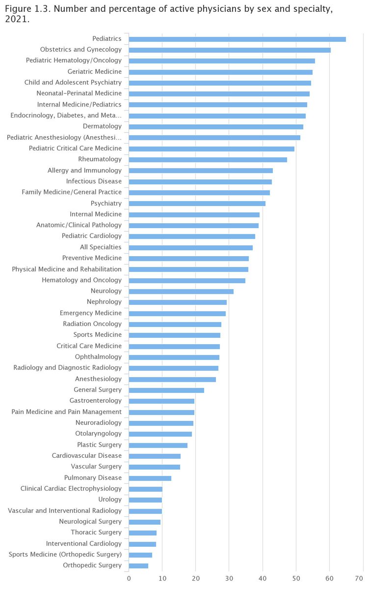
New generation of US #doctors show progress, by @aamc study on residents and fellows, 47% total female, #gensurg 46%, #radiology still only 27%, #emed 39%…
Based on the @StanfordMed graphic, we can assume why progress for gensurg but why not for Rads…
Based on the @StanfordMed graphic, we can assume why progress for gensurg but why not for Rads…

♂ 50 y. With pulsatile femoral mass. history of femoral puncture 💉.
1/4
#vasculardoppler #ultrasound #MedEd #FOAMed #POCUS #radiology @ButterflyNetInc
1/4
#vasculardoppler #ultrasound #MedEd #FOAMed #POCUS #radiology @ButterflyNetInc
2/4
Dx?
Dx?
3/4 Ying-yang , or pepsi sign described in both true and false aneurysms.
On Doppler ultrasound, the yin-yang sign indicates bidirectional flow due to the swirling of blood within the true or false aneurysm. twitter.com/i/web/status/1…
On Doppler ultrasound, the yin-yang sign indicates bidirectional flow due to the swirling of blood within the true or false aneurysm. twitter.com/i/web/status/1…

A 57-YO Mexican♀️, works on a farm has several pets, including dogs, at home: epigastric fullness, and a 15-pound weight loss.
CT: complex cystic left liver mass.
1/5
doi.org/10.1093/cid/ci…
#gastroenterology #radiology #medicine
CT: complex cystic left liver mass.
1/5
doi.org/10.1093/cid/ci…
#gastroenterology #radiology #medicine

Resection: Cyst wall (red➡️), daughter cysts (*) normal liver tissue (yellow⬅️)
🔬acellular laminated wall (*), inner germinal layer (black⬅️), & protoscoleces surrounded by a broad capsule (green⬅️). Refractile hooklets (blue⬅️) & calcareous bodies (red⬅️)
2/5
#GIPath #IDtwitter

🔬acellular laminated wall (*), inner germinal layer (black⬅️), & protoscoleces surrounded by a broad capsule (green⬅️). Refractile hooklets (blue⬅️) & calcareous bodies (red⬅️)
2/5
#GIPath #IDtwitter
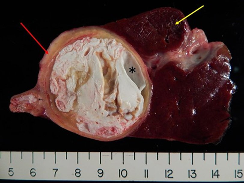
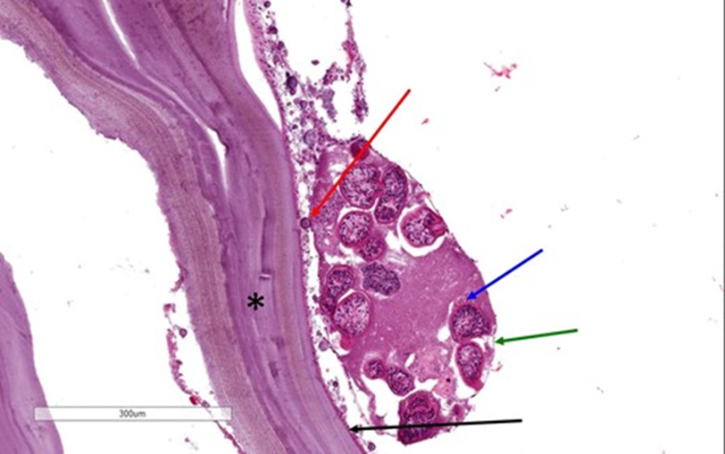
ECHINOCOCCAL CYST
The patient underwent total excision of the cyst followed by albendazole therapy for 4 weeks.
3/5
doi.org/10.1093/cid/ci…
#microbiology #GITwitter #MedStudentTwitter
The patient underwent total excision of the cyst followed by albendazole therapy for 4 weeks.
3/5
doi.org/10.1093/cid/ci…
#microbiology #GITwitter #MedStudentTwitter
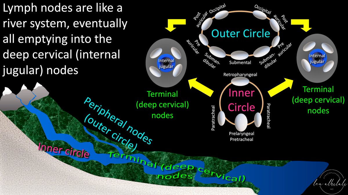
1/To be or not 2b?? That is the question!
Do you have questions about how to remember cervical lymph node anatomy & levels?
Here’s a #tweetorial to show you how--#Superbowl weekend edition!
#medtwitter #meded #neurorad #HNrad #FOAMed #FOAMrad #radres #radtwitter #ENT #radiology
Do you have questions about how to remember cervical lymph node anatomy & levels?
Here’s a #tweetorial to show you how--#Superbowl weekend edition!
#medtwitter #meded #neurorad #HNrad #FOAMed #FOAMrad #radres #radtwitter #ENT #radiology

A 17-YO: a 3-week of dizziness, headache, & weakness of the R leg
MR: ring-enhancing lesions in the frontal lobe & basal ganglia on the L side (A; T1-W) as well as surrounding edema & midline shift (B; T2-W)
1/6
DOI: 10.1056/NEJMicm2202196
#neurology #radiology #IDTwitter
MR: ring-enhancing lesions in the frontal lobe & basal ganglia on the L side (A; T1-W) as well as surrounding edema & midline shift (B; T2-W)
1/6
DOI: 10.1056/NEJMicm2202196
#neurology #radiology #IDTwitter

IgG ab for Echinococcus multilocularis & E. granulosus: ➖
Surgical excision🔬: necrotic tissue, granulomatous inflammation, echinococcal laminated membrane (C, arrow) & an intact cyst (D, arrow)
RT-PCR: ➕for E. multilocularis
2/6
#parasitology #Pathologists #pathology
Surgical excision🔬: necrotic tissue, granulomatous inflammation, echinococcal laminated membrane (C, arrow) & an intact cyst (D, arrow)
RT-PCR: ➕for E. multilocularis
2/6
#parasitology #Pathologists #pathology

CT imaging of the chest and abdomen did not show any other sites of disease.
CEREBRAL ALVEOLAR ECHINOCOCCOSIS
3/6
DOI: 10.1056/NEJMicm2202196
#microbiology #MedTwitter #Doctor
CEREBRAL ALVEOLAR ECHINOCOCCOSIS
3/6
DOI: 10.1056/NEJMicm2202196
#microbiology #MedTwitter #Doctor
A 14-yo ♂️ lived on a farm: a 1-month history of episodic headaches, vomiting, & papilledema
MR: a multiloculated cyst of the brain (A) with a hypointense rim and small projections in T2 phase (B, arrow)
1/5
DOI: 10.1056/NEJMicm2208104
#neurology #radiology #pediatric
MR: a multiloculated cyst of the brain (A) with a hypointense rim and small projections in T2 phase (B, arrow)
1/5
DOI: 10.1056/NEJMicm2208104
#neurology #radiology #pediatric
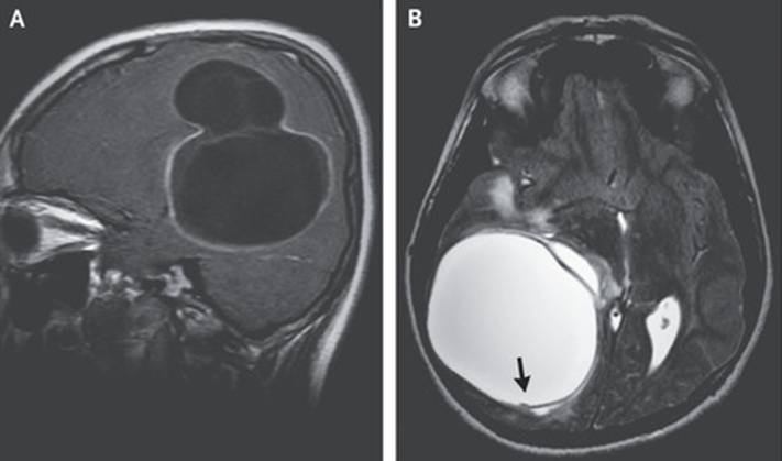
Findings suggestive of CYSTIC ECHINOCOCCOSIS.
CT of the body: no other sites of disease.
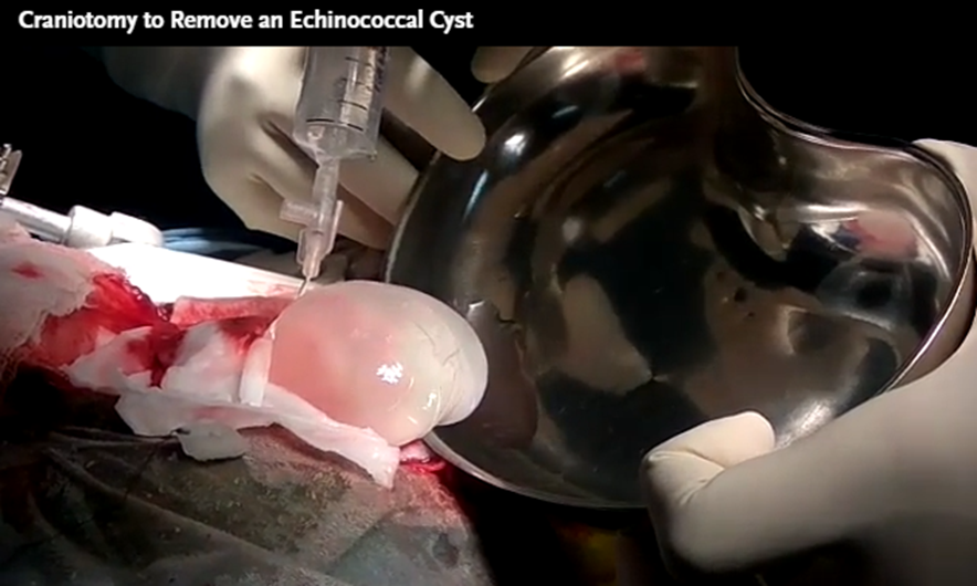
A craniotomy was performed, during which saline irrigation was used to separate the cyst wall from the brain to avoid rupture.
2/5
DOI: 10.1056/NEJMicm2208104
#IDtwitter #parasitology
CT of the body: no other sites of disease.
A craniotomy was performed, during which saline irrigation was used to separate the cyst wall from the brain to avoid rupture.
2/5
DOI: 10.1056/NEJMicm2208104
#IDtwitter #parasitology
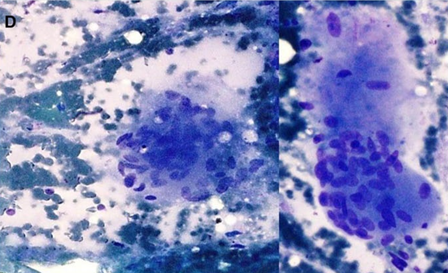
🔬: an echinococcal laminated membrane lined by a germinal layer with daughter cysts (Panel D, arrows) and protoscolices (inset, arrows) with hooklets (arrowhead).
PRIMARY CEREBRAL CYSTIC ECHINOCOCCOSIS FROM ECHINOCOCCUS GRANULOSUS
3/5
#microbiology #Pathologists #pathology
PRIMARY CEREBRAL CYSTIC ECHINOCOCCOSIS FROM ECHINOCOCCUS GRANULOSUS
3/5
#microbiology #Pathologists #pathology

Super easy explain of physiologic Hepatic Pulsed Wave Doppler waveform.
🧵1/
#doppler #radiology #pocus #vexus #hemodinamics #butterflyiq @ButterflyNetInc @hepocus
🧵1/
#doppler #radiology #pocus #vexus #hemodinamics #butterflyiq @ButterflyNetInc @hepocus
Lets understand the basic principles of chest x-ray interpretation (1/10)
Use the inside-out approach. Start from inside most structures and then go outward (2/10)
Look at trachea, mediastinum, cardiac size & borders. Don't forget looking at the hilum (3/10)
A 32-YO♀️ from Guatemala, Ph+ B-ALL post #chemotherapy: #dyspnea
CT: ground-glass opacities (2A yellow box), interstitial pulmonary edema with septal thickening (2B yellow circles), & pericardial effusion (2C yellow arrows), lymph nodes (2D yellow arrows)
Eosinophilia
1/7
CT: ground-glass opacities (2A yellow box), interstitial pulmonary edema with septal thickening (2B yellow circles), & pericardial effusion (2C yellow arrows), lymph nodes (2D yellow arrows)
Eosinophilia
1/7

Due to normal ejection fraction, the differential diagnosis of dyspnea included non-cardiogenic pulmonary edema, pneumonitis secondary to chemotoxicity, and infection.
2/7
doi.org/10.1016/j.idcr…
#haematology #radiology #IDtwitter
2/7
doi.org/10.1016/j.idcr…
#haematology #radiology #IDtwitter

She progressed to acute hypoxic respiratory failure.
🔬bronchoalveolar lavage: numerous larvae (3A,3B) with short buccal grove (arrow head) and prominent genital primordium (arrow) consistent with STRONGYLOIDES HYPERINFECTION
3/7
#lungdisease #parasitology #Pathologists
🔬bronchoalveolar lavage: numerous larvae (3A,3B) with short buccal grove (arrow head) and prominent genital primordium (arrow) consistent with STRONGYLOIDES HYPERINFECTION
3/7
#lungdisease #parasitology #Pathologists

A 4-YO♂️ treated for tuberculosis for 3 years with chronic cough, kyphosis, recurrent pneumonia, rib osteomyelitis: lung consolidation, mediastinal lymphadenopathy, cervical kyphosis; R-sided paravertebral soft tissue swelling (T3 to T6)
1/7
#IDtwitter #radiology #Orthopedics
1/7
#IDtwitter #radiology #Orthopedics

Serology: ➕for Aspergillus flavus & Aspergillus fumigatus.
Aspiration of the paravertebral soft tissue:
🔬granulomatous inflammation
🧫Acinetobacter spp.
VERTEBRAL OSTEOMYELITIS & ACINETOBACTER SPP PARAVERTEBRAL SOFT TISSUE INFECTION
2/7
#microbiology #MedTwitter
Aspiration of the paravertebral soft tissue:
🔬granulomatous inflammation
🧫Acinetobacter spp.
VERTEBRAL OSTEOMYELITIS & ACINETOBACTER SPP PARAVERTEBRAL SOFT TISSUE INFECTION
2/7
#microbiology #MedTwitter
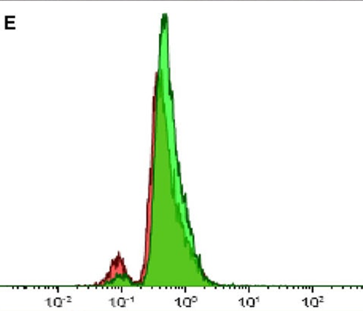
Nitroblue tetrazolium dye reduction test: no reduction of the nitroblue tetrazolium
No right shift noted in the activated neutrophils in flow cytometry-based DHR assay (E, patient; F, control)
CHRONIC GRANULOMATOUS DISEASE
3/7
DOI: 10.1097/INF.0000000000001221
#medicine

No right shift noted in the activated neutrophils in flow cytometry-based DHR assay (E, patient; F, control)
CHRONIC GRANULOMATOUS DISEASE
3/7
DOI: 10.1097/INF.0000000000001221
#medicine

A 6-YO Bedouin ♂️: a limp, headache, left facial nerve palsy, & left hemiparesis.
Blood eosinophil count of 890/μL.
MRI: a brain cyst.
1/8
DOI: doi.org/10.4269/ajtmh.…
#neurology #pediatric #MedTwitter
Blood eosinophil count of 890/μL.
MRI: a brain cyst.
1/8
DOI: doi.org/10.4269/ajtmh.…
#neurology #pediatric #MedTwitter

Abdominal ultrasound and chest radiography did not reveal additional cysts.
Blood serology (ELISA) for ECHINOCOCCOSIS: ➖
The brain cyst was fully removed surgically.
2/8
DOI: doi.org/10.4269/ajtmh.…
#neuro #Pediatrics #surgery
Blood serology (ELISA) for ECHINOCOCCOSIS: ➖
The brain cyst was fully removed surgically.
2/8
DOI: doi.org/10.4269/ajtmh.…
#neuro #Pediatrics #surgery

Histopathological examination of the brain cyst fluid showed several echinococcal larvae (C and D), confirming CYSTIC ECHOCOCCOSIS diagnosis.
Over the following 4 months, he was treated with albendazole without complications.
3/8
#parasitology #microbiology #IDtwitter
Over the following 4 months, he was treated with albendazole without complications.
3/8
#parasitology #microbiology #IDtwitter

An 80-YO ♂️ diabetes, ischaemic heart disease, & end stage kidney disease requiring haemodialysis:
a radio-opaque nodule in X-ray.
1/2
#radiology #Emergency #healthcare
thelancet.com/article/S0140-…
a radio-opaque nodule in X-ray.
1/2
#radiology #Emergency #healthcare
thelancet.com/article/S0140-…

Continuous glucose monitoring device causes consternation on chest x-ray
2/2
#endocrinology #medicine
doi.org/10.1016/S0140-…
thelancet.com/article/S0140-…
2/2
#endocrinology #medicine
doi.org/10.1016/S0140-…
thelancet.com/article/S0140-…
The radio-opaque object had a complex internal
structure consisting of a microchip, a battery, and an
electronic circuit.
The subcutaneous continuous glucose monitoring
transmitter device had been displaced from deltoid muscle to the patient’s back.
#MedicalStudents
structure consisting of a microchip, a battery, and an
electronic circuit.
The subcutaneous continuous glucose monitoring
transmitter device had been displaced from deltoid muscle to the patient’s back.
#MedicalStudents
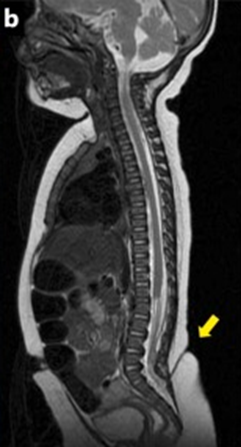
A 3-month-old ♂️, a dimple at the center of a lumbosacral hemangioma (a): fever & deteriorating condition.
CSF: ⬆️cell count 5717/µL;⬆️protein = 352 mg/dL; ⬇️glucose < 10 mg/dL.
RM (b): ?
1/7
doi.org/10.1016/j.idcr…
#pediatric #IDtwitter #Neurology
CSF: ⬆️cell count 5717/µL;⬆️protein = 352 mg/dL; ⬇️glucose < 10 mg/dL.
RM (b): ?
1/7
doi.org/10.1016/j.idcr…
#pediatric #IDtwitter #Neurology

RM b): a congenital dermal sinus communicating with the spinal cord.
Enterobacter aerogenes was isolated from CSF and stool cultures
CONGENITAL DERMAL SINUS ASSOCIATED WITH MENINGITIS BY ENTEROBACTER AEROGENES
2/7
#radiology #microbiology #pediatria
Enterobacter aerogenes was isolated from CSF and stool cultures
CONGENITAL DERMAL SINUS ASSOCIATED WITH MENINGITIS BY ENTEROBACTER AEROGENES
2/7
#radiology #microbiology #pediatria
Congenital dermal sinus is associated with meningitis caused by atypical pathogens.
Enterobacter aerogenes community-acquired infections associated with congenital dermal sinus are rarely observed.
3/7
#bacteriology #MedTwitter
Enterobacter aerogenes community-acquired infections associated with congenital dermal sinus are rarely observed.
3/7
#bacteriology #MedTwitter
Resources to study #Neurology
Adams and Victor’s. By far my favourite textbook.
The book is more clinically oriented, coloured with anecdotes and mental models. Reading it feels like seeing a pt in the ward/OPD
#MedTwitter #neurotwitter
Adams and Victor’s. By far my favourite textbook.
The book is more clinically oriented, coloured with anecdotes and mental models. Reading it feels like seeing a pt in the ward/OPD
#MedTwitter #neurotwitter

It has elements of philosophy, history and is written eloquently.
Whimsical, yet profound, it’s teachings stay with me. Added bonus, my Guru in Neurology finds mention in the text 🙃 I’d recommend this for #mbbs #md and #dm students

Whimsical, yet profound, it’s teachings stay with me. Added bonus, my Guru in Neurology finds mention in the text 🙃 I’d recommend this for #mbbs #md and #dm students


Bradley is the standard #textbook in #neurology. A great book, it’s more like #Harrison. Great for information, latest research and management. Essential for the DM #neurology candidate, but also useful for MD #internalmedicine 

80 yo ♀️ with chronic cough.
What would you do with this "ugly" lung lesion? 🧵👇🏻
#radres #radtwitter #radiology #chestrad
What would you do with this "ugly" lung lesion? 🧵👇🏻
#radres #radtwitter #radiology #chestrad

❓❓❓
A 65-YO man, from North African: asymptomatic dorsolumbar mass for 30 years with normal skin appearance.
MRI: ?
1/6
doi.org/10.1093/cid/ci…
#radiology #MedTwitter #parasitology

MRI: ?
1/6
doi.org/10.1093/cid/ci…
#radiology #MedTwitter #parasitology

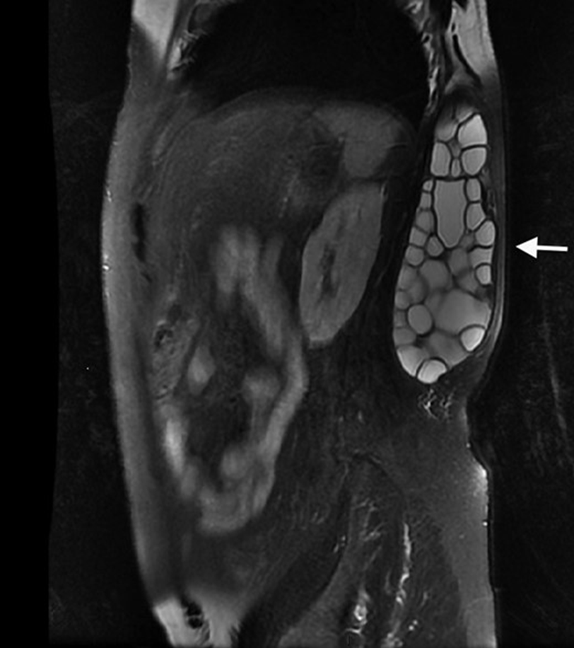
MRI: a multivesicular cyst in the latissimus dorsi muscle (arrow)
🧪for Echinococcus granulosus: ELISA, & Western blot➕
Surgery: multiple daughter vesicles of E. granulosus, with the presence of multiple scolices
INTRAMUSCULAR HYDATID CYST
2/6
#parasites #medicine #radres

🧪for Echinococcus granulosus: ELISA, & Western blot➕
Surgery: multiple daughter vesicles of E. granulosus, with the presence of multiple scolices
INTRAMUSCULAR HYDATID CYST
2/6
#parasites #medicine #radres


No puncture should be performed, to avoid dissemination of the cysts that can cause an anaphylactic shock.
The patient received 3 other courses of albendazole: serologic control became negative after 1 year.
3/6
#IDtwitter #surgery
The patient received 3 other courses of albendazole: serologic control became negative after 1 year.
3/6
#IDtwitter #surgery
HYPOTHALAMUS (HT)🧵-the control center of circadian rhythm, fatigue/wakefulness, hunger/satiety, sex drive, thirst/BP—and the command post for endocrine control via the HT-pituitary axis.
#meded #neuroradiology #neuroscience #radiology #neurology #neurosurgery #neuroanatomy
1/28
#meded #neuroradiology #neuroscience #radiology #neurology #neurosurgery #neuroanatomy
1/28
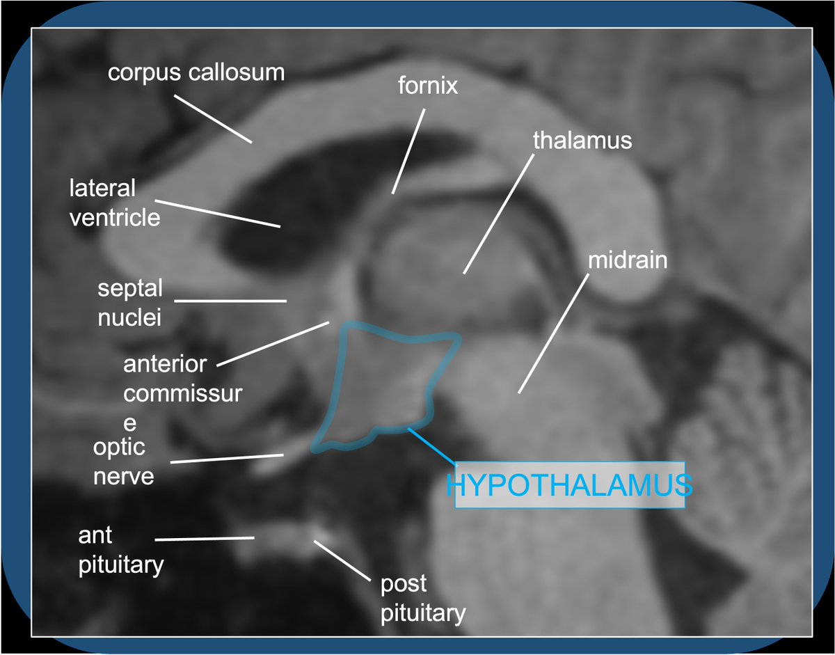
Quiz A: Histamine activity in the brain/brainstem contributes to alertness/wakefulness–hence: sleepy effects of benadryl. Histaminergic neurosecretory cell bodies are exclusively in the tuberomammillary nucleus of the TUBER CINEREUM (HT floor), with widespread projections.
2/28
2/28

The HT is a mysterious and complex—I get confused sighs from medical students when the topic arises—the small almond-sized morsel is the control center for endocrine/hormone regulation, and homeostasis of food/water consumption, sleep, BP, sex/attachment. The HT is boss!
3/28
3/28
A 59-YO DM, steroid, myasthenia gravis with thymoma: headache & deviation of tongue to the R side with fasciculation suggestive of right 12th cranial nerve palsy
CT: sphenoid sclerotic changes, soft tissue density lesion with calcification.
1/9
#neurology #radiology #IDtwitter
CT: sphenoid sclerotic changes, soft tissue density lesion with calcification.
1/9
#neurology #radiology #IDtwitter

MRI brain: a hypointense lesion involving the body of sphenoid (R > L) and clivus on T2 weighted images and T1 weighted images with moderate contrast enhancement possibly fungal in aetiology.
2/9
doi.org/10.1016/j.idcr…
#radiologist #radres #MedTwitter

2/9
doi.org/10.1016/j.idcr…
#radiologist #radres #MedTwitter


Endoscopic sphenoidotomy & debridement: invasive fungal sphenoid sinusitis & CENTRAL SKULL BASE OSTEOMYELITIS involving the clivus
🔬from sphenoid mucosa: morphologically ASPERGILLUS with foci of tissue invasion.
3/9
#microbiology #maxillofacial #Orthopedics
🔬from sphenoid mucosa: morphologically ASPERGILLUS with foci of tissue invasion.
3/9
#microbiology #maxillofacial #Orthopedics
🎉Introducing RoentGen, a generative vision-language foundation model based on #StableDiffusion, fine-tuned on a large chest x-ray and radiology report dataset, and controllable through text prompts!
@PierreChambon6 @Dr_ASChaudhari @curtlanglotz
🧵#Radiology #AI #StanfordAIMI
@PierreChambon6 @Dr_ASChaudhari @curtlanglotz
🧵#Radiology #AI #StanfordAIMI
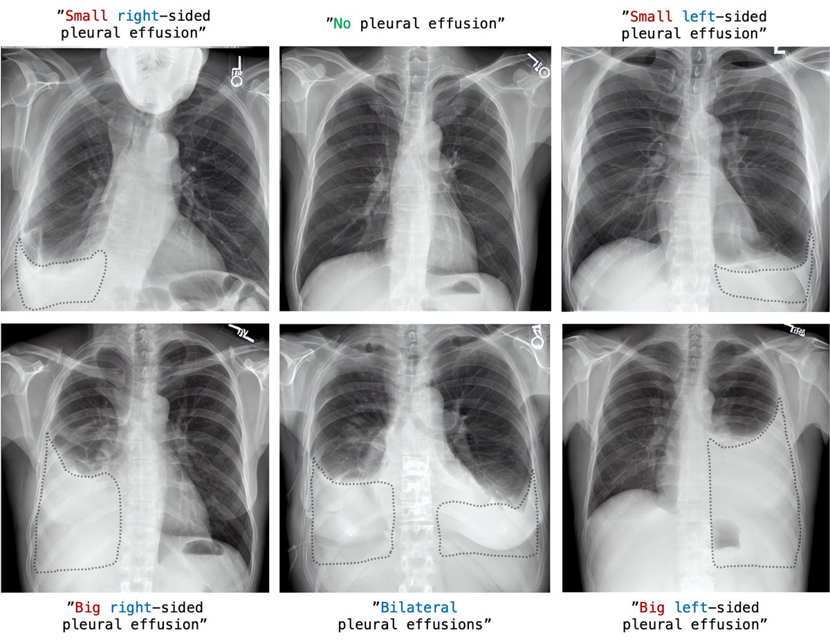
#RoentGen is able to generate a wide variety of radiological chest x-ray (CXR) findings with fidelity and high level of detail. Of note, this is without being explicitly trained on class labels. 

Built on previous work, #RoentGen is a fine-tuned latent diffusion model based on #StableDiffusion. Free-form medical text prompts are used to condition a denoising process, resulting in high-fidelity yet diverse CXR, improving on a typical limitation of GAN-based methods. 

1/They say form follows function! Brain #MRI anatomy is best understood in terms of both form & function
A #tweetorial on how to remember important functional #brain #anatomy
#meded #medtwitter #neurosurgery #neurology #neurorad #FOAMed #FOAMrad #radiology #medstudent #radres
A #tweetorial on how to remember important functional #brain #anatomy
#meded #medtwitter #neurosurgery #neurology #neurorad #FOAMed #FOAMrad #radiology #medstudent #radres


