Discover and read the best of Twitter Threads about #neurorad
Most recents (24)
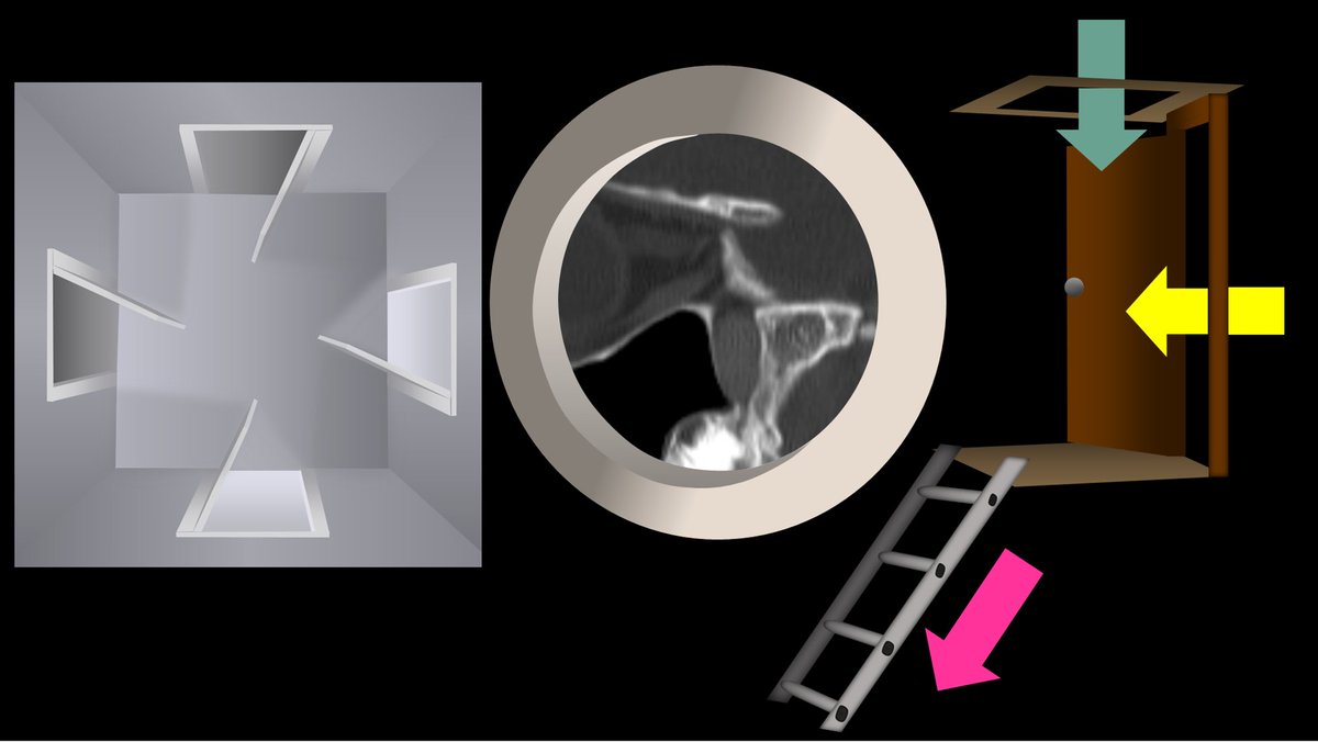
1/Does PTERYGOPALATINE FOSSA anatomy feel as confusing as its spelling? Does it seem to have as many openings as letters in its name?
Let this #tweetorial on PPF #anatomy help you out
#meded #medtwitter #FOAMed #FOAMrad #neurosurgery #neurology #neurorad #neurotwitter #radres
Let this #tweetorial on PPF #anatomy help you out
#meded #medtwitter #FOAMed #FOAMrad #neurosurgery #neurology #neurorad #neurotwitter #radres
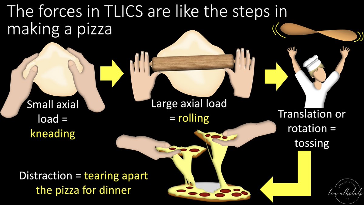
1/Remembering spinal fracture classifications is back breaking work!
A #tweetorial to help your remember the scoring system for thoracic & lumbar fractures—“TLICS” to the cool kids!
#medtwitter #radres #FOAMed #FOAMrad #neurorad #Meded #backpain #spine #Neurosurgery
A #tweetorial to help your remember the scoring system for thoracic & lumbar fractures—“TLICS” to the cool kids!
#medtwitter #radres #FOAMed #FOAMrad #neurorad #Meded #backpain #spine #Neurosurgery

1/Understanding cervical radiculopathy is a pain in the neck! But knowing the distributions can help your search
A #tweetorial to help you remember cervical radicular pain distributions
#medtwitter #radres #FOAMed #FOAMrad #neurorad #Meded #meded #spine #Neurosurgery
A #tweetorial to help you remember cervical radicular pain distributions
#medtwitter #radres #FOAMed #FOAMrad #neurorad #Meded #meded #spine #Neurosurgery

1/ “Say Aaaaaaah!” I was explaining to my resident how I remember tongue anatomy on imaging & he said, “You have to put it on Twitter!”
So here's a #tweetorial about how to remember tongue anatomy on imaging.
#medtwitter #radres #medstudent #FOAMed #FOAMrad #neurorad #meded
So here's a #tweetorial about how to remember tongue anatomy on imaging.
#medtwitter #radres #medstudent #FOAMed #FOAMrad #neurorad #meded
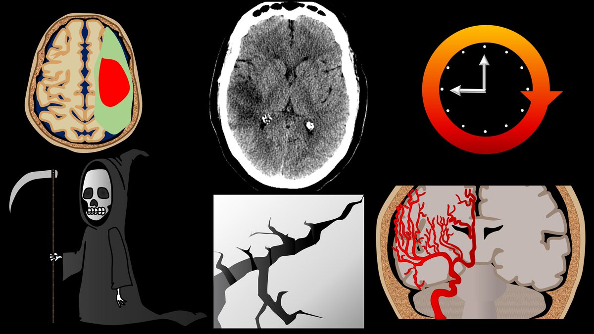
1/Time is brain! So you don’t have time to struggle w/that stroke alert head CT.
Here’s a #tweetorial to help you with the CT findings in acute stroke.
#medtwitter #FOAMed #FOAMrad #ESOC #medstudent #neurorad #radres #meded #radtwitter #stroke #neurology #neurotwitter
Here’s a #tweetorial to help you with the CT findings in acute stroke.
#medtwitter #FOAMed #FOAMrad #ESOC #medstudent #neurorad #radres #meded #radtwitter #stroke #neurology #neurotwitter
Interesting case in this patient with acute right-sided weakness
#neurorad #neurotwitter #meded #Neurosurgery #Neurology @TheASNR @RSNA #medtwitter


#neurorad #neurotwitter #meded #Neurosurgery #Neurology @TheASNR @RSNA #medtwitter

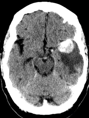
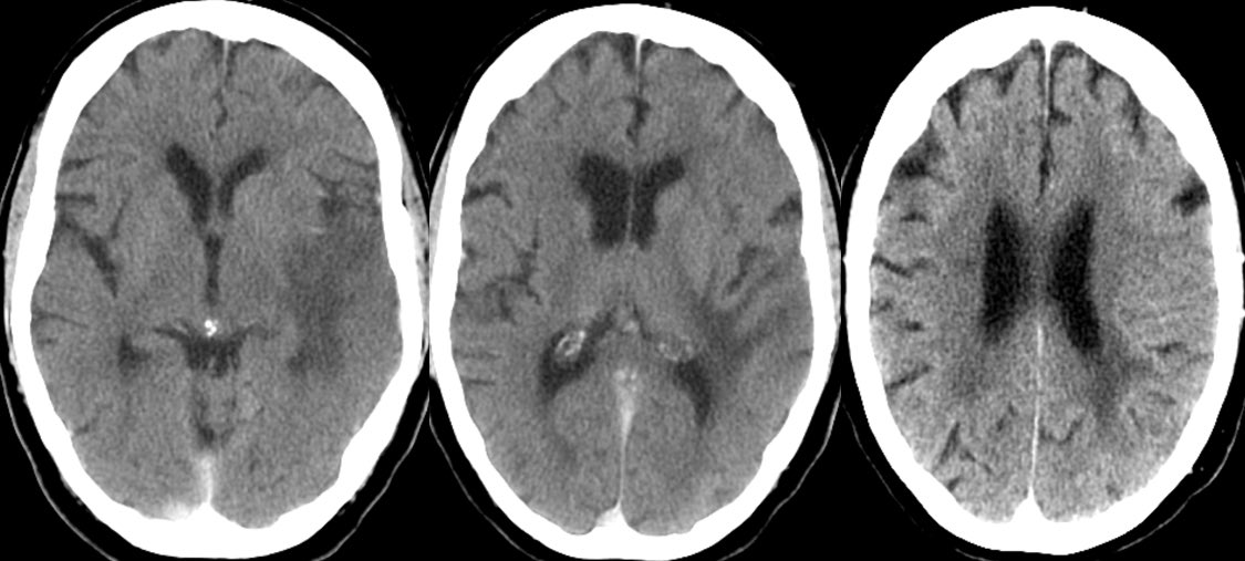

Can you determine the diagnosis off the CT?
▶️Initial non-con CT shows a 3cm hyperdense lobulated extra-axial mass in the expected region of the left MCA bifurcation, consistent with a giant aneurysm. There are associated peripheral calcifications
▶️ What is the cause of the surrounding hypodensity?
▶️ What is the cause of the surrounding hypodensity?
1/”Tell me where it hurts.” How back pain radiates can tell you where the lesion is—if you know where to look!
A #tweetorial about how to remember lumbar radicular pain distributions.
#medstudenttwitter #medtwitter #radres #FOAMed #FOAMrad #neurorad #tweetorial #Meded
A #tweetorial about how to remember lumbar radicular pain distributions.
#medstudenttwitter #medtwitter #radres #FOAMed #FOAMrad #neurorad #tweetorial #Meded

Learning case in this 40 y/o F with history of whole brain radiation as a child for brain tumor treatment
#NeuroRad #neurosurgery #Neurology @TheASNR #NeuroTwitter #meded #radres


#NeuroRad #neurosurgery #Neurology @TheASNR #NeuroTwitter #meded #radres


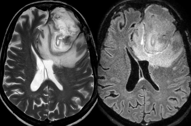
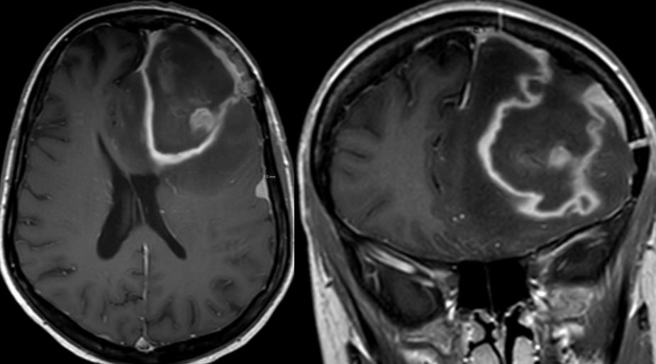
We were consulted for a 2nd opinion.
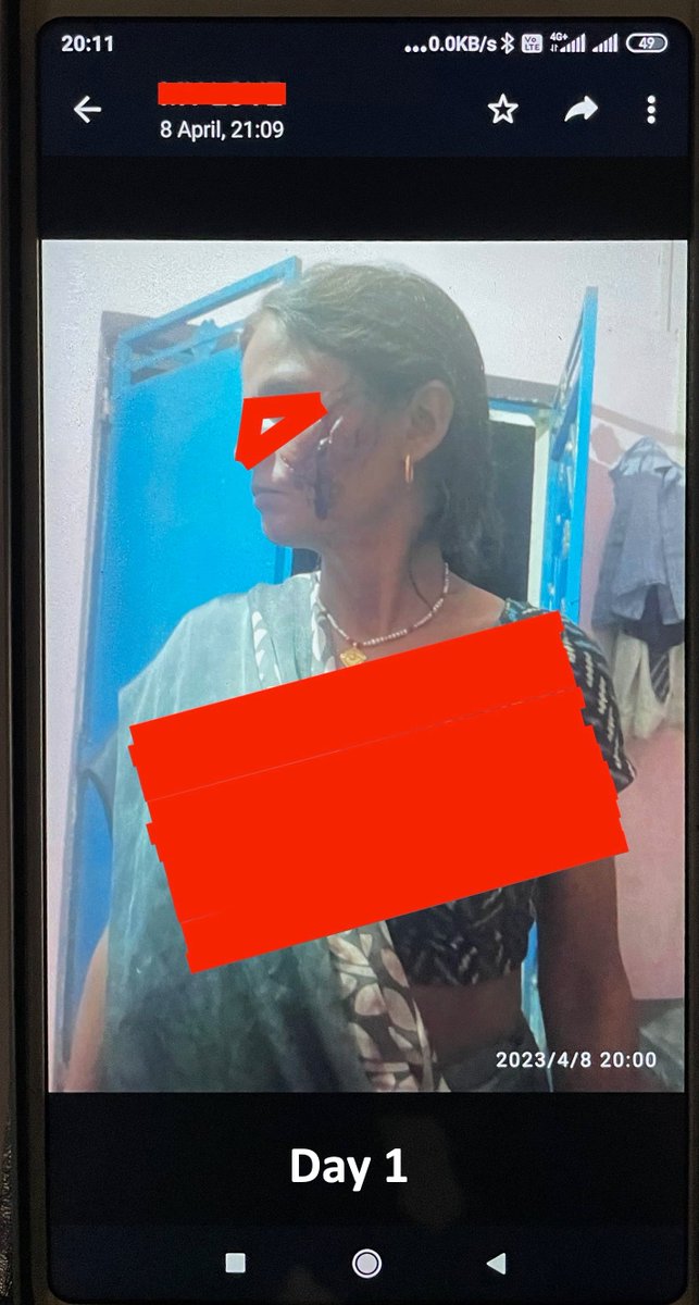
30F. D1. Deep dog bite L cheek while tending fields. Multiple others bitten. Dog was killed the same day by villagers. #NeuroTwitter #MedTwitter #IDtwitter #NeuroRad #Neurology
30F. D1. Deep dog bite L cheek while tending fields. Multiple others bitten. Dog was killed the same day by villagers. #NeuroTwitter #MedTwitter #IDtwitter #NeuroRad #Neurology
1/Do radiologists sound like they are speaking a different language when they talk about MRI? T1 shortening what? T2 prolongation who?
Here’s a translation w/a #tweetorial introduction to MRI.
#medtwitter #FOAMed #FOAMrad #medstudent #neurorad #radres #ASNR23 #neurosurgery
Here’s a translation w/a #tweetorial introduction to MRI.
#medtwitter #FOAMed #FOAMrad #medstudent #neurorad #radres #ASNR23 #neurosurgery

1/Don’t let all your effort be in VEIN!
Developmental venous anomalies (DVAs) are often thought incidental but ignore them at your own risk!
A #tweetorial about how to know when DVAs are the most important finding
#meded #medtwitter #neurorad #neurotwitter #radtwitter #radres
Developmental venous anomalies (DVAs) are often thought incidental but ignore them at your own risk!
A #tweetorial about how to know when DVAs are the most important finding
#meded #medtwitter #neurorad #neurotwitter #radtwitter #radres

ASNR COTW #143
Hx: Altered mental status with oral pain.
NO SPOILERS!!! Give hints in the form of GIF or answer attached poll.
Answer in 24 hrs
#Neuro #Neurorad #Erad #radres #FOAMed #FOAMrad #medtwitter #ASNRCOTW
Hx: Altered mental status with oral pain.
NO SPOILERS!!! Give hints in the form of GIF or answer attached poll.
Answer in 24 hrs
#Neuro #Neurorad #Erad #radres #FOAMed #FOAMrad #medtwitter #ASNRCOTW
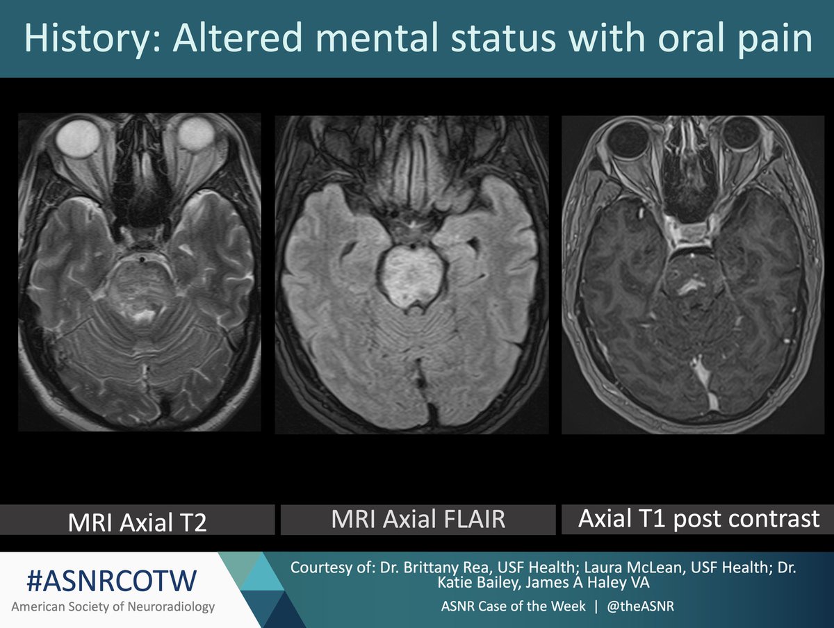
What disease process is associated with oral ulcers and enhancing hyperintense T2 signal lesions in the brain and spinal cord?
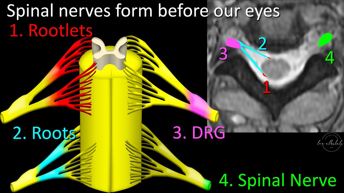
1/Is trying to understand peripheral nerve injury getting on your last nerve? Is the brachial plexus breaking you?
Here’s a #tweetorial to help you understand, recognize & remember the classification of peripheral nerve injuries
#medtwitter #meded #FOAMed #neurorad #neurotwitter
Here’s a #tweetorial to help you understand, recognize & remember the classification of peripheral nerve injuries
#medtwitter #meded #FOAMed #neurorad #neurotwitter

ASNR COTW #142
Hx: 62 y/o male initially found unresponsive on the street presents with bradycardia and anterograde amnesia.
NO SPOILERS!!! Give hints in the form of GIF or answer attached poll.
Answer in 24 hrs
#Neuro #Neurorad #radres #FOAMed #FOAMrad #medtwitter #ASNRCOTW
Hx: 62 y/o male initially found unresponsive on the street presents with bradycardia and anterograde amnesia.
NO SPOILERS!!! Give hints in the form of GIF or answer attached poll.
Answer in 24 hrs
#Neuro #Neurorad #radres #FOAMed #FOAMrad #medtwitter #ASNRCOTW

What is the most likely diagnosis given the history and MRI findings in the referenced images?
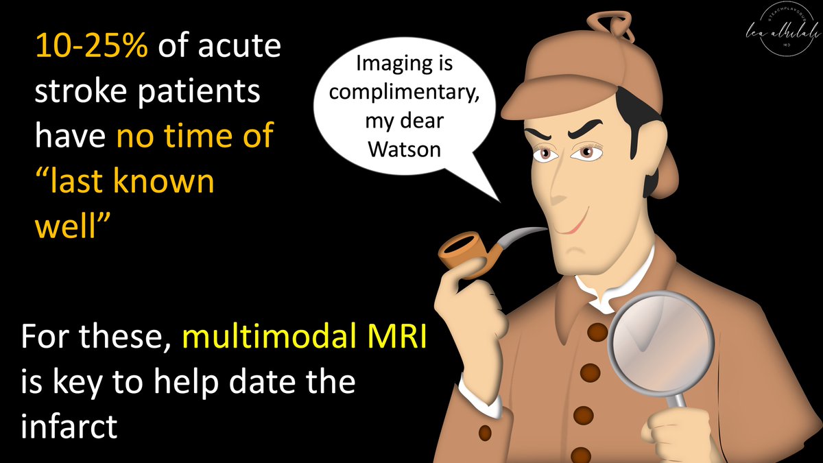
1/Time is brain! But what time is it?
If you don’t know the time of stroke onset, are you able to deduce it from imaging?
Here’s a #tweetorial to help you date a #stroke on MR!
#medtwitter #meded #neurotwitter #neurology #neurorad #radres #radtwitter #radiology #FOAMed #FOAMrad
If you don’t know the time of stroke onset, are you able to deduce it from imaging?
Here’s a #tweetorial to help you date a #stroke on MR!
#medtwitter #meded #neurotwitter #neurology #neurorad #radres #radtwitter #radiology #FOAMed #FOAMrad
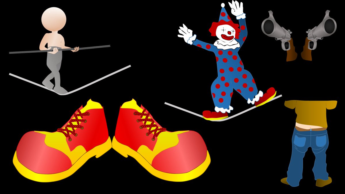
1/Feeling unarmed when it comes to evaluating cervical radiculopathy & foraminal narrowing on MR?
Here’s a #tweetorial that’ll take that weight off your shoulder & show you how to rate cervical foraminal stenosis!
#medtwitter #meded #FOAMed #radtwitter #neurorad #spine #radres
Here’s a #tweetorial that’ll take that weight off your shoulder & show you how to rate cervical foraminal stenosis!
#medtwitter #meded #FOAMed #radtwitter #neurorad #spine #radres

1/Does the work up for dizziness make your head spin?
Wondering what you should look for on an MRI for dizziness?
Here’s a #tweetorial on what you can (and can’t) see on MRI in #dizziness
#medtwitter #meded #neurotwitter #neurorad #radres #HNrad #neurotwitter #stroke #FOAMed
Wondering what you should look for on an MRI for dizziness?
Here’s a #tweetorial on what you can (and can’t) see on MRI in #dizziness
#medtwitter #meded #neurotwitter #neurorad #radres #HNrad #neurotwitter #stroke #FOAMed

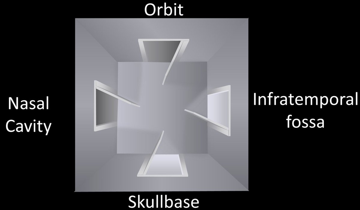
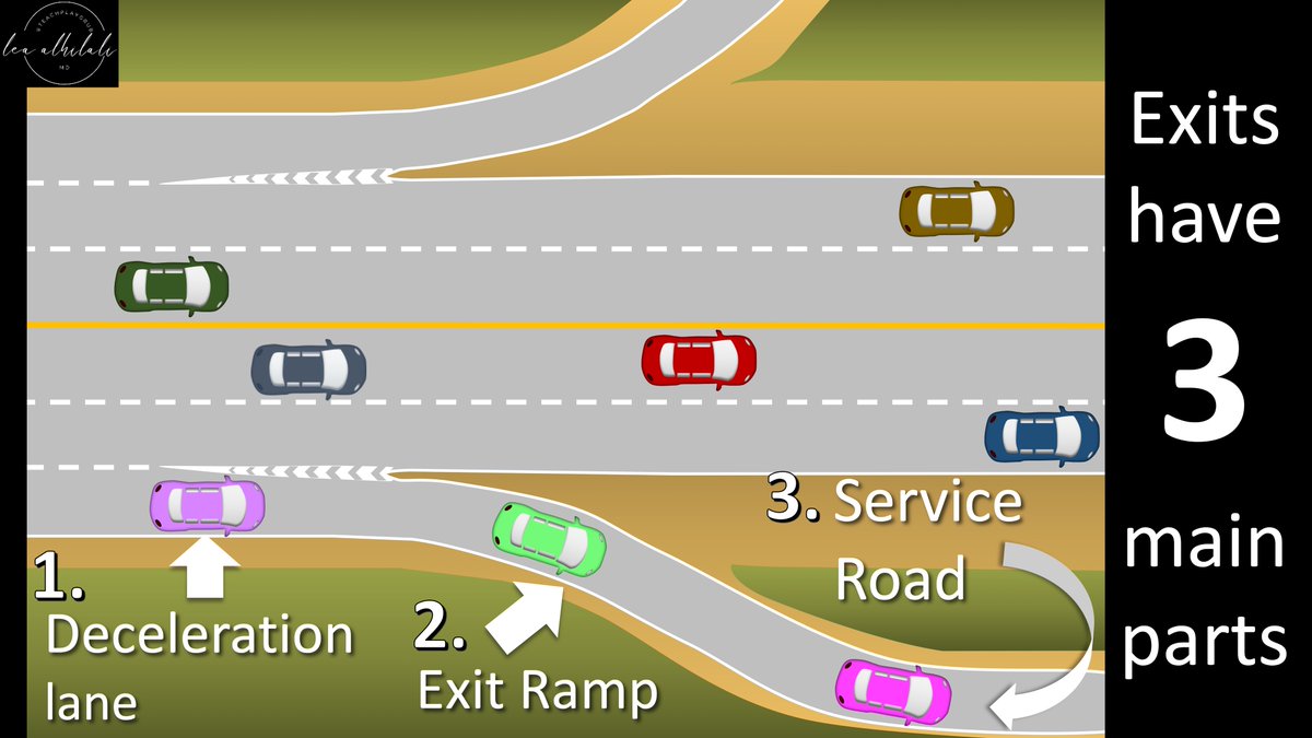
1/I call the skullbase “homebase” bc you can’t make an anatomy homerun without it!
Most know the arteries of the skullbase, but few know the veins. Do you?
Here’s a🧵to help you remember #skullbase venous #anatomy!
#medtwitter #meded #neurorad #radtwitter #neurosurgery #radres
Most know the arteries of the skullbase, but few know the veins. Do you?
Here’s a🧵to help you remember #skullbase venous #anatomy!
#medtwitter #meded #neurorad #radtwitter #neurosurgery #radres

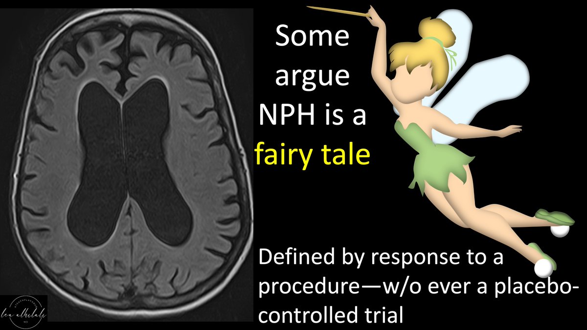
1/To call it or not to call it? That is the question!
Do you feel a bit wacky & wobbly when it comes to calling normal pressure hydrocephalus on imaging?
Here’s a #tweetorial about imaging NPH!
#medtwitter #meded #neurotwitter #neurorad #radres #dementia #neurosurgery #FOAMed
Do you feel a bit wacky & wobbly when it comes to calling normal pressure hydrocephalus on imaging?
Here’s a #tweetorial about imaging NPH!
#medtwitter #meded #neurotwitter #neurorad #radres #dementia #neurosurgery #FOAMed

1/I always say you can tell a bad read on a spine MR if it doesn’t talk about the lateral recess
What will I think when I see your read? What do you say about lateral recesses?
Here’s a #tweetorial on lateral recess #anatomy & grading stenosis
#medtwitter #neurorad #spine
What will I think when I see your read? What do you say about lateral recesses?
Here’s a #tweetorial on lateral recess #anatomy & grading stenosis
#medtwitter #neurorad #spine

1/Does trying to remember inferior frontal gyrus anatomy leave you speechless?
Do you get a Broca’s aphasia trying to name the parts?
Here’s a #tweetorial to help you remember the #anatomy of this important region
#medtwitter #meded #neurotwitter #neurorad #radtwitter #radres
Do you get a Broca’s aphasia trying to name the parts?
Here’s a #tweetorial to help you remember the #anatomy of this important region
#medtwitter #meded #neurotwitter #neurorad #radtwitter #radres

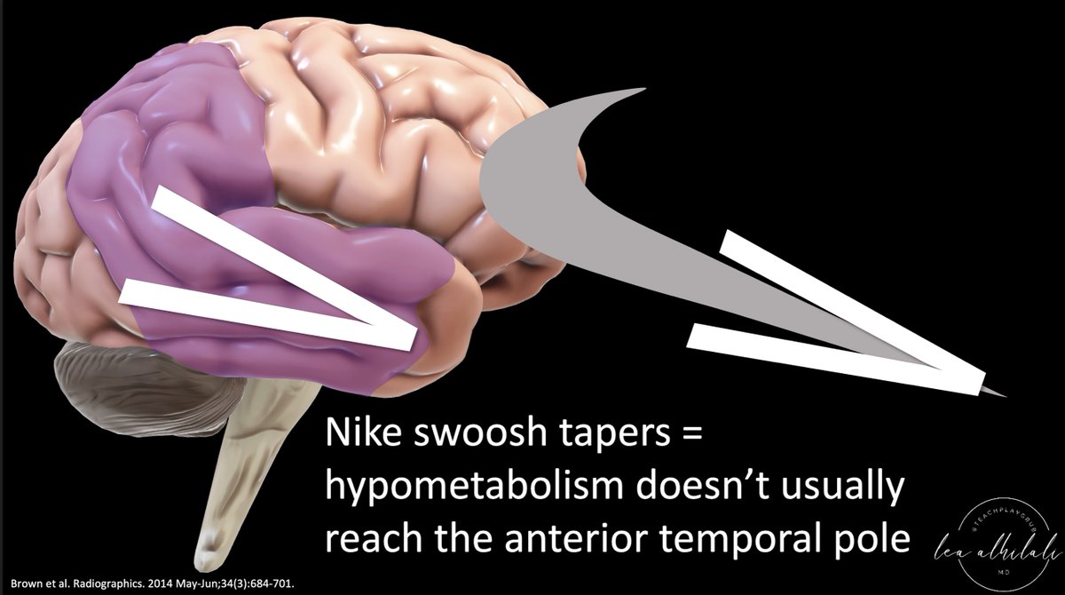
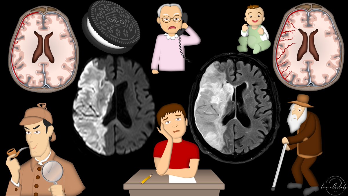
1/Having trouble remembering how to differentiate dementias on imaging?
Here’s a #tweetorial to show you how to remember the imaging findings in dementia & never forget!
#medtwitter #meded #neurorad #radres #dementia #alzheimers #neurotwitter #neurology #FOAMed #FOAMrad #PET
Here’s a #tweetorial to show you how to remember the imaging findings in dementia & never forget!
#medtwitter #meded #neurorad #radres #dementia #alzheimers #neurotwitter #neurology #FOAMed #FOAMrad #PET

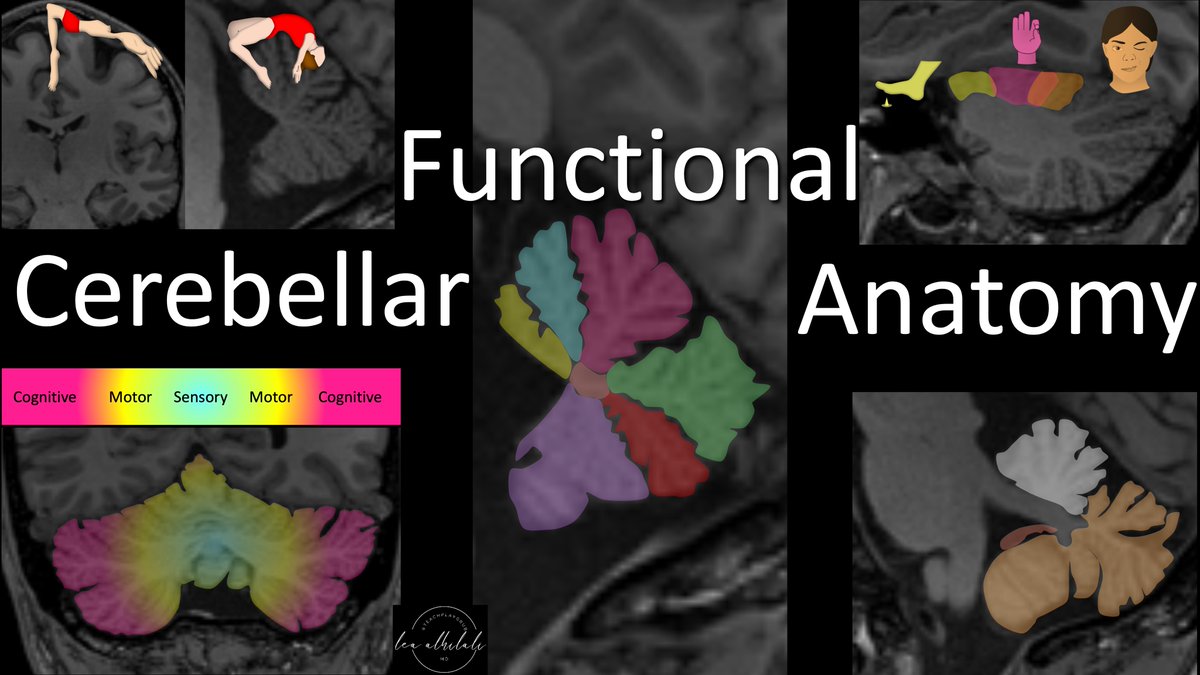
1/Your baby’s all grown up! Cerebellum may mean “little cerebrum” but its jobs are anything but little
Do you know cerebellar anatomy beyond vermis & hemispheres?
Here’s a #tweetorial about the functional #anatomy of the cerebellum!
#medtwitter #neurotwitter #neurorad #meded
Do you know cerebellar anatomy beyond vermis & hemispheres?
Here’s a #tweetorial about the functional #anatomy of the cerebellum!
#medtwitter #neurotwitter #neurorad #meded
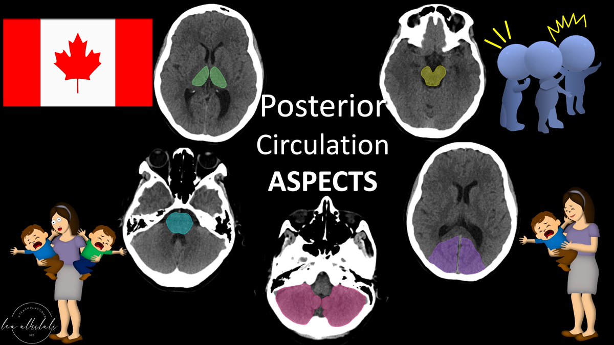
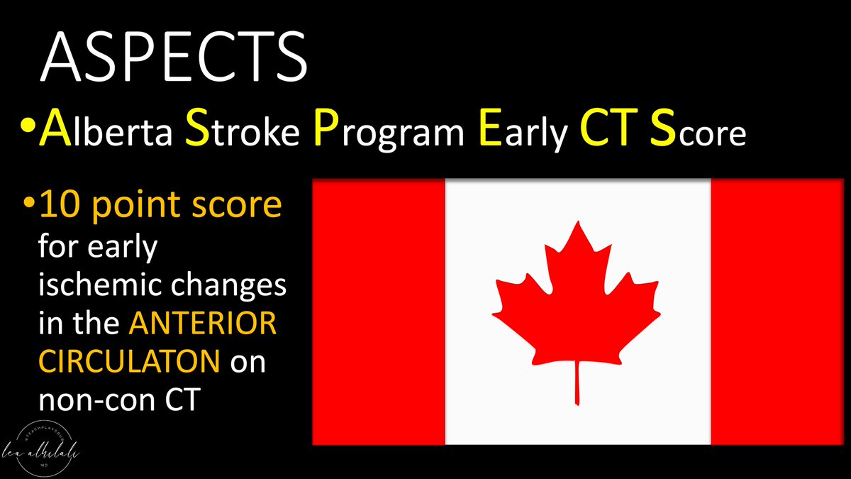
1/Do you know all the aspects of, well, ASPECTS?
Many know the anterior circulation #stroke scoring system—but posterior circulation (pc) ASPECTS is often unknown
Here’s a #tweetorial to help you remember pc-ASPECTS
#medtwitter #neurotwitter #meded #neurorad #neurology #FOAMed
Many know the anterior circulation #stroke scoring system—but posterior circulation (pc) ASPECTS is often unknown
Here’s a #tweetorial to help you remember pc-ASPECTS
#medtwitter #neurotwitter #meded #neurorad #neurology #FOAMed
