Discover and read the best of Twitter Threads about #neurosurgery
Most recents (24)
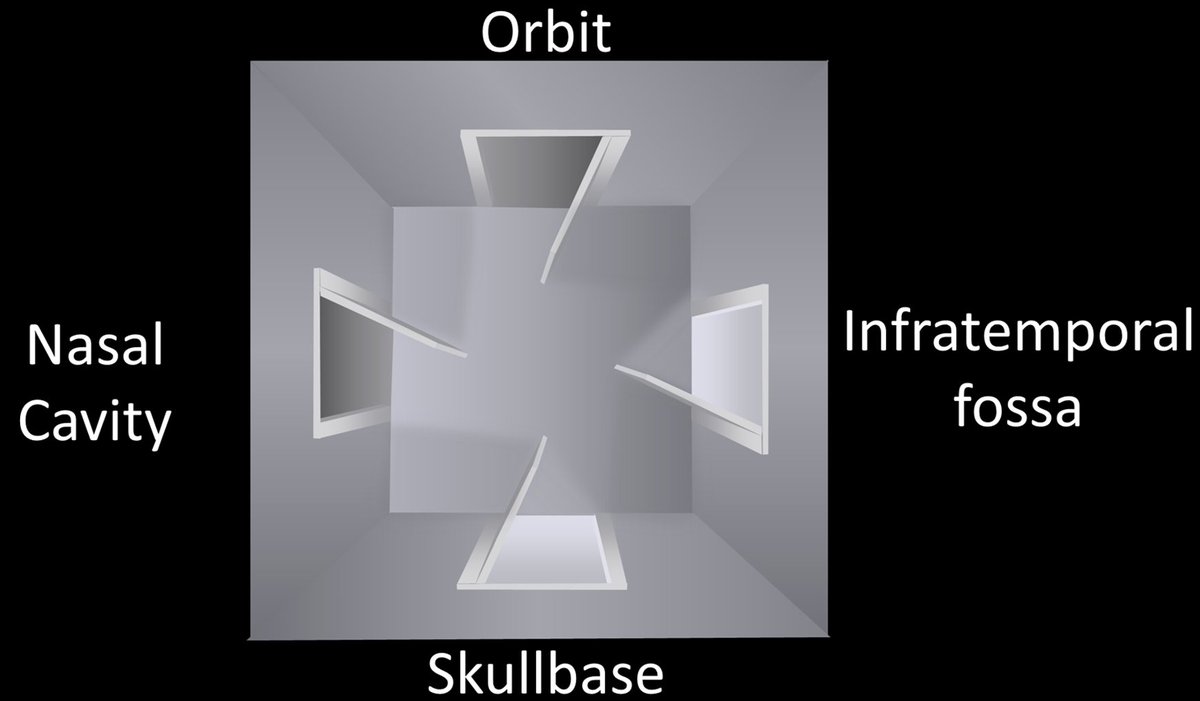
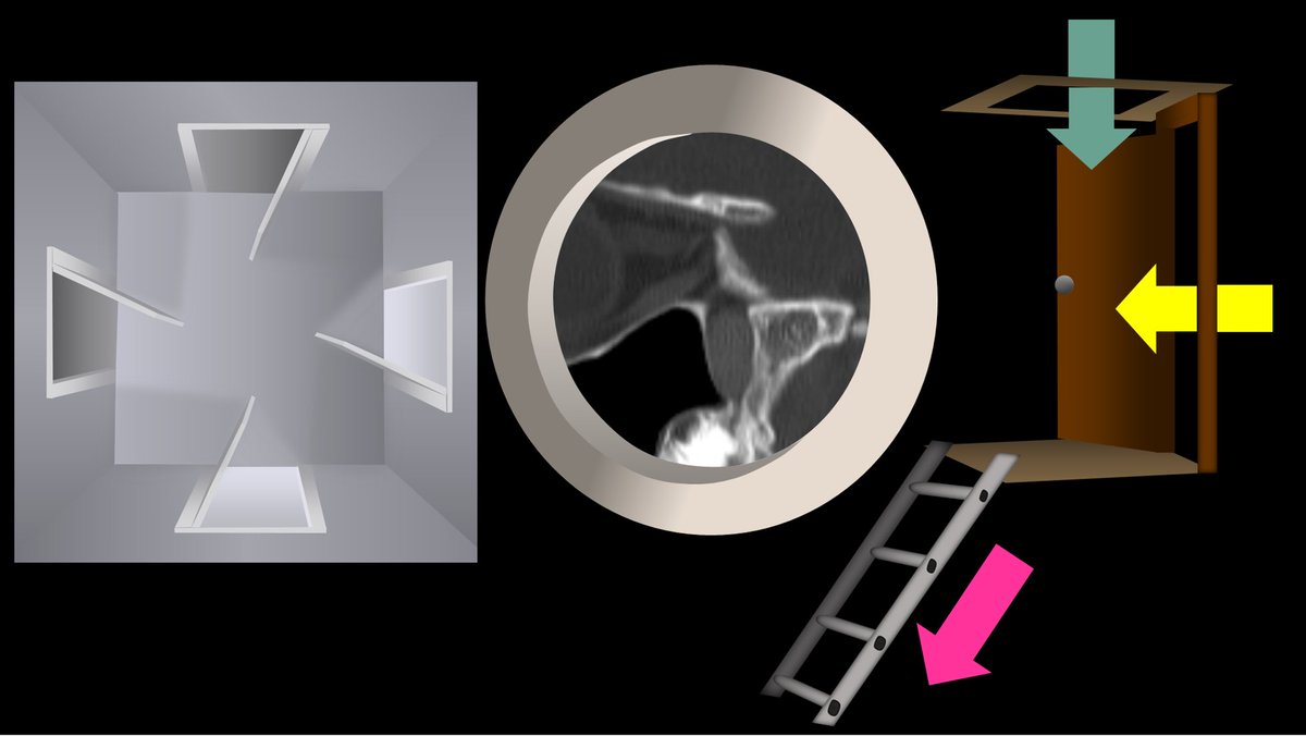
1/Does PTERYGOPALATINE FOSSA anatomy feel as confusing as its spelling? Does it seem to have as many openings as letters in its name?
Let this #tweetorial on PPF #anatomy help you out
#meded #medtwitter #FOAMed #FOAMrad #neurosurgery #neurology #neurorad #neurotwitter #radres
Let this #tweetorial on PPF #anatomy help you out
#meded #medtwitter #FOAMed #FOAMrad #neurosurgery #neurology #neurorad #neurotwitter #radres
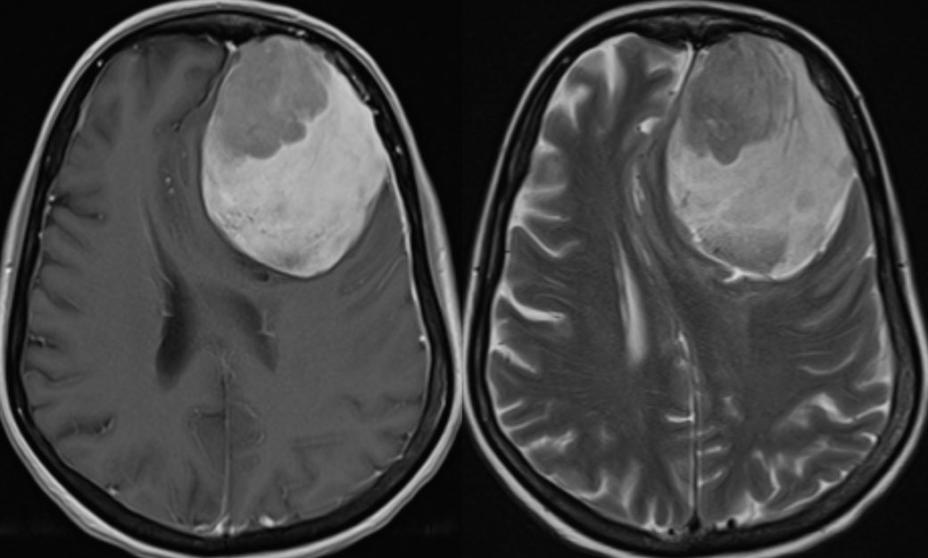
Differential Diagnosis for cortically based masses
P-DOG 🐶
1️⃣Pleomorphic Xanthoastrocytoma (PXA)
2️⃣Dysembryoplastic neuroepithelial tumor (DNET)
3️⃣Oligodendroglioma
4️⃣Ganglioglioma
#Neurology #neurosurgery #peds #radres #neurotwitter @The_ASPNR @TheASNR #MedTwitter


P-DOG 🐶
1️⃣Pleomorphic Xanthoastrocytoma (PXA)
2️⃣Dysembryoplastic neuroepithelial tumor (DNET)
3️⃣Oligodendroglioma
4️⃣Ganglioglioma
#Neurology #neurosurgery #peds #radres #neurotwitter @The_ASPNR @TheASNR #MedTwitter


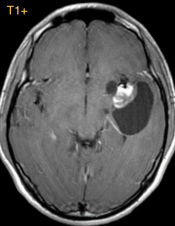
1️⃣PXA
Originate in the subpial astrocytes typically in children and young adults often with a seizure history
Temporal lobe is most common
Originate in the subpial astrocytes typically in children and young adults often with a seizure history
Temporal lobe is most common
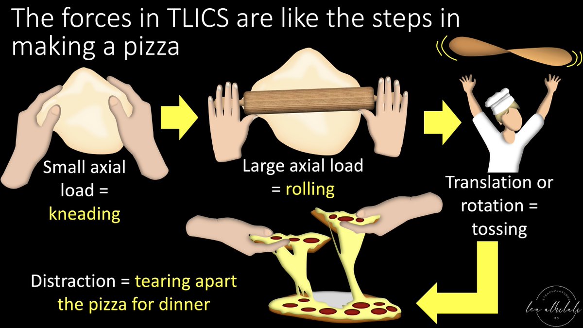
1/Remembering spinal fracture classifications is back breaking work!
A #tweetorial to help your remember the scoring system for thoracic & lumbar fractures—“TLICS” to the cool kids!
#medtwitter #radres #FOAMed #FOAMrad #neurorad #Meded #backpain #spine #Neurosurgery
A #tweetorial to help your remember the scoring system for thoracic & lumbar fractures—“TLICS” to the cool kids!
#medtwitter #radres #FOAMed #FOAMrad #neurorad #Meded #backpain #spine #Neurosurgery

What is the most likely diagnosis in this 30 y/o w/ history of discitis/osteomyelitis presenting w/ fevers, chills, and neck pain? 🧠
#ent #Neurosurgery #Neurology #medtwitter #MedEd @The_ASSR #NeuroTwitter


#ent #Neurosurgery #Neurology #medtwitter #MedEd @The_ASSR #NeuroTwitter

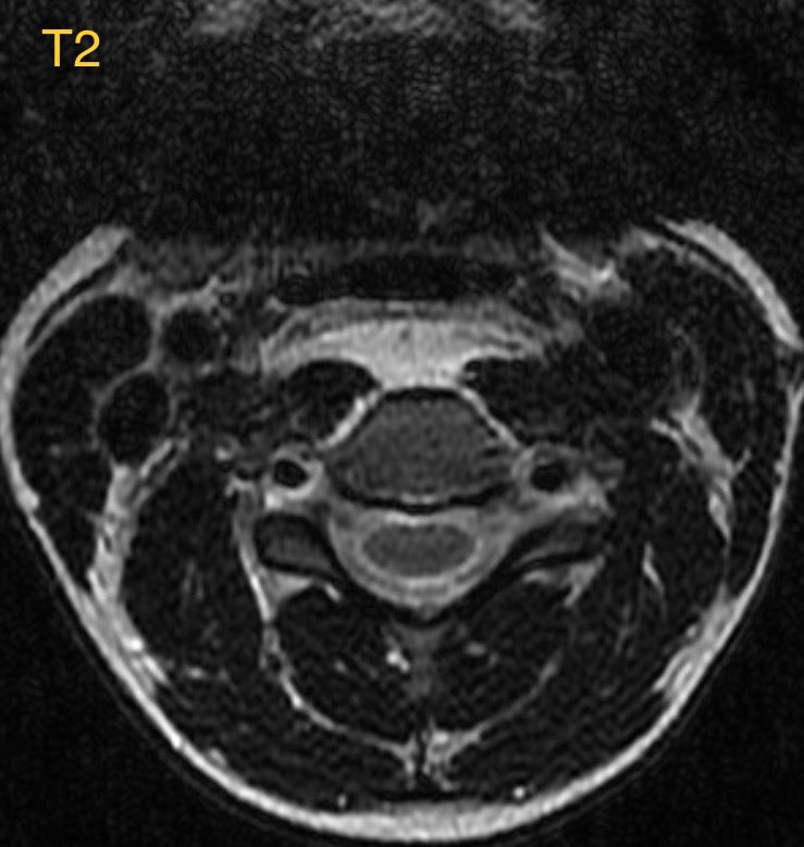


Answer: Longus Colli Calcific Tendinitis
▶️Etiology: inflammatory reaction in response to deposition of calcium hydroxyapatite crystals (just like in the rotator cuff)
▶️This case is a bit tricky as the history is somewhat misleading (though it often is in radiology)
▶️Etiology: inflammatory reaction in response to deposition of calcium hydroxyapatite crystals (just like in the rotator cuff)
▶️This case is a bit tricky as the history is somewhat misleading (though it often is in radiology)
Interesting case, what is the most likely diagnosis in this 25 y/o F w/ 1 year history of migraine headaches, left hand numbness, and b/l retinal artery occlusions? 🧠 👁️
#Ophthalmology #neurology #neurosurgery #neurotwitter #MedEd @TheASNR #MedTwitter


#Ophthalmology #neurology #neurosurgery #neurotwitter #MedEd @TheASNR #MedTwitter


Answer: Susac syndrome 🧠
▶️Susac syndrome is a microangiopathy (likely autoimmune affecting the precapillary arterioles) with a strong female predilection, typically occurring in women age 20-40
▶️Susac syndrome is a microangiopathy (likely autoimmune affecting the precapillary arterioles) with a strong female predilection, typically occurring in women age 20-40
Clinical presentation:
Classic triad
1️⃣Encephalopathy
2️⃣Branch retinal artery occlusions
3️⃣Hearing loss
💡Though most patients do not present with the complete triad (it may develop over years)
Classic triad
1️⃣Encephalopathy
2️⃣Branch retinal artery occlusions
3️⃣Hearing loss
💡Though most patients do not present with the complete triad (it may develop over years)
1/Understanding cervical radiculopathy is a pain in the neck! But knowing the distributions can help your search
A #tweetorial to help you remember cervical radicular pain distributions
#medtwitter #radres #FOAMed #FOAMrad #neurorad #Meded #meded #spine #Neurosurgery
A #tweetorial to help you remember cervical radicular pain distributions
#medtwitter #radres #FOAMed #FOAMrad #neurorad #Meded #meded #spine #Neurosurgery

Tips & tricks of DWI to help narrow the differential
Ddx:
Stroke
Abscess
Hypercellular tumor
Hematoma
Epidermoid cyst
Encephalitis
Seizure
Demyelination
Toxic/metabolic disorders
CJD
Other stuff I’m forgetting
#Neurology #neurosurgery #radres #MedTwitter #MedEd @TheASNR


Ddx:
Stroke
Abscess
Hypercellular tumor
Hematoma
Epidermoid cyst
Encephalitis
Seizure
Demyelination
Toxic/metabolic disorders
CJD
Other stuff I’m forgetting
#Neurology #neurosurgery #radres #MedTwitter #MedEd @TheASNR


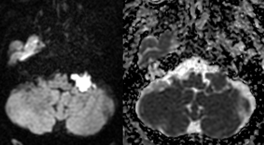
Child with a history of dental caries presents with a firm mass at the angle of the mandible. What is the most likely diagnosis? 🤔 🧠
#neurotwitter #ent #peds #Neurology #neurosurgery @ASHNRSociety @The_ASPNR #MedTwitter


#neurotwitter #ent #peds #Neurology #neurosurgery @ASHNRSociety @The_ASPNR #MedTwitter


Answer: Sclerosing osteomyelitis of Garré
▶️Biopsy showed a reactive and reparative osseous process and bone culture grew oral flora (though cultures are usually negative)
▶️Biopsy showed a reactive and reparative osseous process and bone culture grew oral flora (though cultures are usually negative)
▶️SOG is thought to be due to a low grade infection possibly 2/2 dental disease. However, there should be no signs of acute infection (suppuration, bony sequestration or draining tracts)
Interesting case in this patient with acute right-sided weakness
#neurorad #neurotwitter #meded #Neurosurgery #Neurology @TheASNR @RSNA #medtwitter


#neurorad #neurotwitter #meded #Neurosurgery #Neurology @TheASNR @RSNA #medtwitter

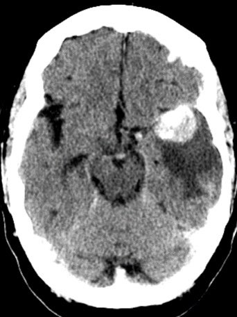
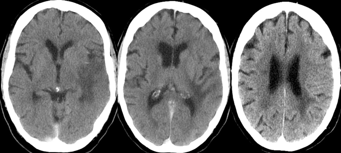

Can you determine the diagnosis off the CT?
▶️Initial non-con CT shows a 3cm hyperdense lobulated extra-axial mass in the expected region of the left MCA bifurcation, consistent with a giant aneurysm. There are associated peripheral calcifications
▶️ What is the cause of the surrounding hypodensity?
▶️ What is the cause of the surrounding hypodensity?
Case of emphysematous epiglottitis in an adult
Epiglottitis is an emergency as it can potentially cause airway compromise especially in children who have smaller airways #neurotwitter #ent #peds #Neurosurgery #MedTwitter #MedEd @ASHNRSociety

Epiglottitis is an emergency as it can potentially cause airway compromise especially in children who have smaller airways #neurotwitter #ent #peds #Neurosurgery #MedTwitter #MedEd @ASHNRSociety


▶️In children the diagnosis can be confirmed with upright plain film. CT requires placing the patient supine which may exacerbate inspiratory strider
▶️In adults, the diagnosis may not be suspected clinically so patients may end up with a CT scan as in this case
▶️In adults, the diagnosis may not be suspected clinically so patients may end up with a CT scan as in this case
▶️Bacterial infection typically 2/2 H. Influenza in unvaccinated children
▶️In adults, possible pathogens include Strep, Staph, and H. influ
▶️In adults, possible pathogens include Strep, Staph, and H. influ
Case of diffuse CSF seeding of tumor in this patient w/ WHO grade 4 diffuse hemispheric glioma
#NeuroTwitter #neurosurgery #Neurology #peds #futureradres @The_ASPNR #MedEd


#NeuroTwitter #neurosurgery #Neurology #peds #futureradres @The_ASPNR #MedEd


▶️Prospectively this mass was thought to be an embryonal tumor w/ multilayered rosettes given the marked diffusion restriction, hemorrhage, and lack of surrounding edema 🧠
Learning case in this 40 y/o F with history of whole brain radiation as a child for brain tumor treatment
#NeuroRad #neurosurgery #Neurology @TheASNR #NeuroTwitter #meded #radres


#NeuroRad #neurosurgery #Neurology @TheASNR #NeuroTwitter #meded #radres


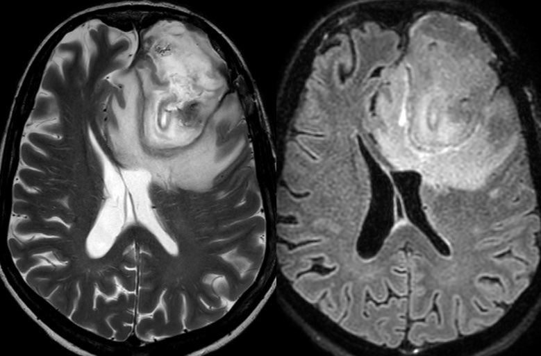
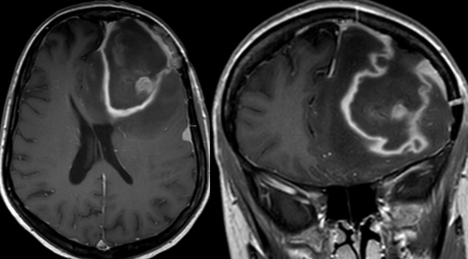
Interesting case of complicated acute bacterial rhinosinusitis in this child with no PMH presenting w/ HA, fever & L sided weakness
#NeuroTwitter #ent #radres #neurosurgery @TheASNR @ASHNRSociety @PhilipRChapman1 #radres #futureradres


#NeuroTwitter #ent #radres #neurosurgery @TheASNR @ASHNRSociety @PhilipRChapman1 #radres #futureradres


1/Do you want a BASIC approach to skullBASE lesions?
My FINAL tweetorial on skullbase lesions—posterior skullbase & overall approach!
This #tweetorial will teach you to diagnose skullbase lesions by answering only TWO simple questions!
#medtwitter #meded #neurosurgery #radres
My FINAL tweetorial on skullbase lesions—posterior skullbase & overall approach!
This #tweetorial will teach you to diagnose skullbase lesions by answering only TWO simple questions!
#medtwitter #meded #neurosurgery #radres

1/Talk about the bases being loaded!
Central skull base has some of the most complicated anatomy & pathology in neuro
Do you know how to approach it?
Here’s a #tweetorial to show you how diagnose lesions at the central skullbase!
#meded #medtwitter #FOAMed #neurosurgery
Central skull base has some of the most complicated anatomy & pathology in neuro
Do you know how to approach it?
Here’s a #tweetorial to show you how diagnose lesions at the central skullbase!
#meded #medtwitter #FOAMed #neurosurgery

1/It’s called the skullBASE but it’s anything but BASIC!
Does the sight of a skullbase lesion strike fear into your heart?
Never fear! Here’s a #tweetorial about a simple approach to these lesions that will change how you look at these cases
#medtwitter #meded #neurosurgery
Does the sight of a skullbase lesion strike fear into your heart?
Never fear! Here’s a #tweetorial about a simple approach to these lesions that will change how you look at these cases
#medtwitter #meded #neurosurgery

1/Do radiologists sound like they are speaking a different language when they talk about MRI? T1 shortening what? T2 prolongation who?
Here’s a translation w/a #tweetorial introduction to MRI.
#medtwitter #FOAMed #FOAMrad #medstudent #neurorad #radres #ASNR23 #neurosurgery
Here’s a translation w/a #tweetorial introduction to MRI.
#medtwitter #FOAMed #FOAMrad #medstudent #neurorad #radres #ASNR23 #neurosurgery

What is the most likely diagnosis in this adolescent with seizure? 🧠
(Sorry I have no CT without)
#neurotwitter #peds #Neurosurgery #Neurology @The_ASPNR @TheASNR #MedTwitter


(Sorry I have no CT without)
#neurotwitter #peds #Neurosurgery #Neurology @The_ASPNR @TheASNR #MedTwitter


What is the most likely diagnosis?
Answer: Confirmed supratentorial ependymoma
Predicting tumors is incredibly challenging in the absence of specific features …some learning points on the case in 🧵
Predicting tumors is incredibly challenging in the absence of specific features …some learning points on the case in 🧵
Case of a radiation induced pseudoaneurysm in this patient with headache and AMS 🧠
Imaging in thread #Neurosurgery #Neurology #neurotwitter #radres #MedEd #MedTwitter @TheASNR


Imaging in thread #Neurosurgery #Neurology #neurotwitter #radres #MedEd #MedTwitter @TheASNR


▶️Initial head CT shows subarachnoid hemorrhage centered in the right cerebellopontine angle cistern
▶️CTA confirms an aneurysm of the right anterior inferior cerebellar artery (AICA)

▶️CTA confirms an aneurysm of the right anterior inferior cerebellar artery (AICA)

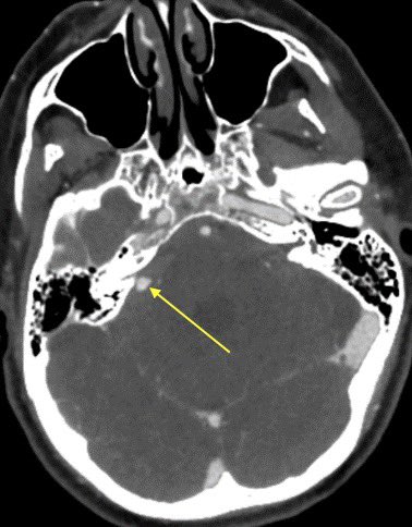
▶️MR displays and ice cream shaped enhancing mass extending through the right internal auditory canal into the cerebellopontine angle cistern, consistent with a vestibular schwannoma #icecream
▶️Careful search into the history confirms the schwannoma was treated with radiation

▶️Careful search into the history confirms the schwannoma was treated with radiation


What is the most likely diagnosis in this 25 y/o M with headache? 🧠
Answer later tonight #radres #Neurology #Neurosurgery #MedEd #MedTwitter #NeuroTwitter @RSNA


Answer later tonight #radres #Neurology #Neurosurgery #MedEd #MedTwitter #NeuroTwitter @RSNA


Most likely diagnosis?
Answer: confirmed germinoma, all these masses are on the differential for a pineal region mass …perhaps the most helpful clue is the age and gender rather than the imaging 🧠
Glioblastoma is the most common variety of astrocytoma
The presence of necrosis is the characteristic feature of glioblastoma
Imaging details in thread #Neurosurgery #neurotwitter #radres #MedTwitter #Neurology @TheASNR


The presence of necrosis is the characteristic feature of glioblastoma
Imaging details in thread #Neurosurgery #neurotwitter #radres #MedTwitter #Neurology @TheASNR


Preoperative approach to sellar region masses, what the surgeon needs to know (at least what I think they need to know)
Additional reporting tips from surgeons are welcomed and encouraged! #Neurosurgery @TheASNR #radres #MedEd #MedTwitter #futureradres #endocrine #Neurology
Additional reporting tips from surgeons are welcomed and encouraged! #Neurosurgery @TheASNR #radres #MedEd #MedTwitter #futureradres #endocrine #Neurology

1/I call the skullbase “homebase” bc you can’t make an anatomy homerun without it!
Most know the arteries of the skullbase, but few know the veins. Do you?
Here’s a🧵to help you remember #skullbase venous #anatomy!
#medtwitter #meded #neurorad #radtwitter #neurosurgery #radres
Most know the arteries of the skullbase, but few know the veins. Do you?
Here’s a🧵to help you remember #skullbase venous #anatomy!
#medtwitter #meded #neurorad #radtwitter #neurosurgery #radres

Can you figure out the cause of hemorrhage in this case?
Imaging and case details in thread #Neurosurgery #radres #MedTwitter #Neurology @TheASNR #MedEd #neurotwitter

Imaging and case details in thread #Neurosurgery #radres #MedTwitter #Neurology @TheASNR #MedEd #neurotwitter


