Discover and read the best of Twitter Threads about #Otolaryngology
Most recents (4)
A 24-YO♀️: a 10-year of episodes of redness & photophobia in eyes, &⬇️visual acuity
SLE: conjunctival hyperemia & peripheral corneal opacification, inflammation & crystalline deposits on the corneal
⬇️
INTERSTITIAL KERATITIS
1/5
DOI: 10.1056/NEJMicm1709103
#eye #internalmedicine
SLE: conjunctival hyperemia & peripheral corneal opacification, inflammation & crystalline deposits on the corneal
⬇️
INTERSTITIAL KERATITIS
1/5
DOI: 10.1056/NEJMicm1709103
#eye #internalmedicine

Etiologies:
✔️Bacterial: SYPHILIS, Lyme borreliosis, tuberculosis, etc.
✔️Viral: Herpesviridae (HERPES SIMPLEX, H. zoster, E-B)...
✔️Parasitic: Onchocerciasis, Acanthamoeba...
✔️Immune: Cogan's syndrome, contact lens-associated keratitis, autoimmune disease…
2/5
#microbiology
✔️Bacterial: SYPHILIS, Lyme borreliosis, tuberculosis, etc.
✔️Viral: Herpesviridae (HERPES SIMPLEX, H. zoster, E-B)...
✔️Parasitic: Onchocerciasis, Acanthamoeba...
✔️Immune: Cogan's syndrome, contact lens-associated keratitis, autoimmune disease…
2/5
#microbiology
ESR:⬆️& evaluation for infectious causes: unremarkable.
6 months after the diagnosis of interstitial keratitis: vertigo, tinnitus, & hearing loss.
Audiometry: sensorineural hearing loss in both ears.
Clinical findings consistent with COGAN´S SYNDROME
3/5
#Ophthalmology #MedEd
6 months after the diagnosis of interstitial keratitis: vertigo, tinnitus, & hearing loss.
Audiometry: sensorineural hearing loss in both ears.
Clinical findings consistent with COGAN´S SYNDROME
3/5
#Ophthalmology #MedEd
A 64-YO ♂️ from a rural community: pain, itching, bleeding in the L ear, & mobile larvae occluding the L external auditory canal
1/4
DOI: 10.1056/NEJMicm2005407
#primarycare #Otolaryngologist
1/4
DOI: 10.1056/NEJMicm2005407
#primarycare #Otolaryngologist

AURAL MYASIS
An ear aspirator, forceps, and irrigation with sterile water were used to remove the larvae
Perforation of the tympanic membrane was identified.
2/4
DOI: 10.1056/NEJMicm2005407
#healthcare #parasitology
An ear aspirator, forceps, and irrigation with sterile water were used to remove the larvae
Perforation of the tympanic membrane was identified.
2/4
DOI: 10.1056/NEJMicm2005407
#healthcare #parasitology

Aural myiasis:
📌infestation of the middle or external ear
📌by fly larvae of the order Diptera.
Risk factors:
✔️chronic otitis media,
✔️diabetes mellitus, and
✔️impaired hygiene.
3/4
DOI: 10.1056/NEJMicm2005407
#Otolaryngology #parasites
📌infestation of the middle or external ear
📌by fly larvae of the order Diptera.
Risk factors:
✔️chronic otitis media,
✔️diabetes mellitus, and
✔️impaired hygiene.
3/4
DOI: 10.1056/NEJMicm2005407
#Otolaryngology #parasites
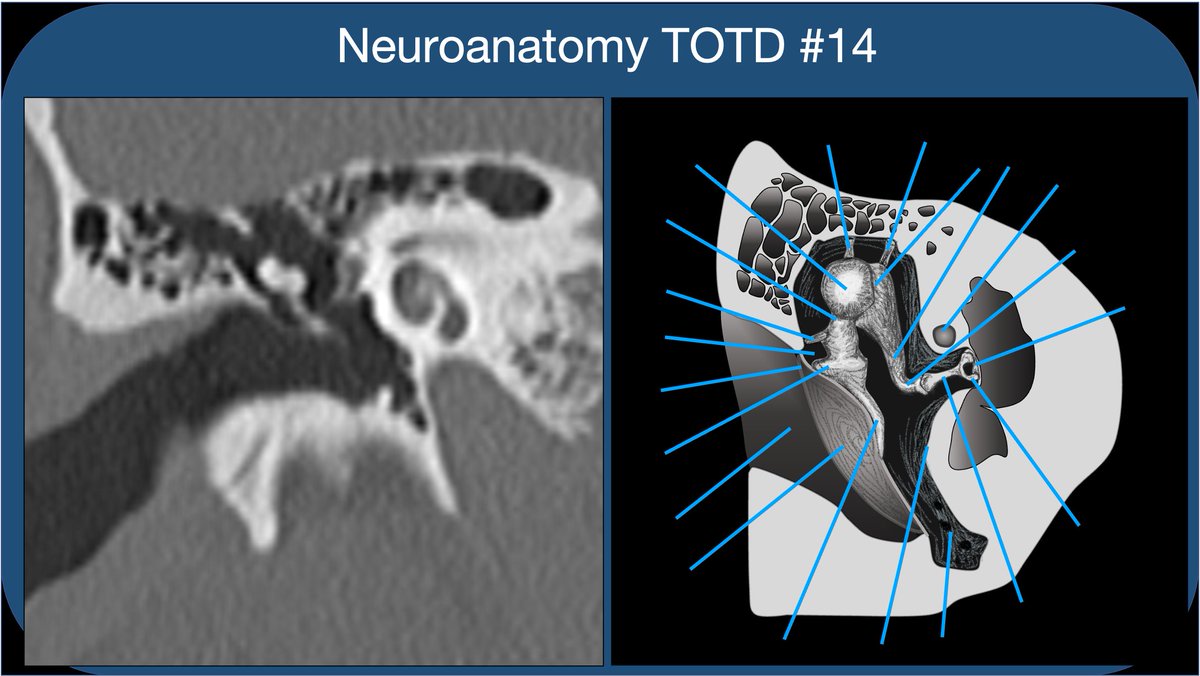
Neuroanatomy TOTD #14🧵
Got some requests to do one of the trickiest areas of human anatomy, the #temporalbone. So many named structures! #meded #FOAMed #FOAMrad #medtwitter #medstudents #radiology #neurorad #radres #neurosurgery #neuroanatomy #ENT #otolaryngology
1/21
Got some requests to do one of the trickiest areas of human anatomy, the #temporalbone. So many named structures! #meded #FOAMed #FOAMrad #medtwitter #medstudents #radiology #neurorad #radres #neurosurgery #neuroanatomy #ENT #otolaryngology
1/21


