Discover and read the best of Twitter Threads about #gipath
Most recents (22)
80 yo M. Known cardiovascular disease and anemia. Acute abdominal pain and vomit.
Diagnosis? Only ONE answer is correct 😉
#radres #futureradres #FOAMrad #FOAMed #GITwitter #Endoscopy #GIpath
1. Ischemic colitis
2. IBD
3. Tumor
4. None of the above
Diagnosis? Only ONE answer is correct 😉
#radres #futureradres #FOAMrad #FOAMed #GITwitter #Endoscopy #GIpath
1. Ischemic colitis
2. IBD
3. Tumor
4. None of the above
A 44-YO♂️, stayed in a rural cottage of France 2 weeks previously: cramping upper abdominal pain with watery diarrhea.
He had passed what he thought were worms in his feces
Eosinophilia
1/6
doi.org/10.1093/cid/ci…
#GITwitter #IDtwitter #microbiology

He had passed what he thought were worms in his feces
Eosinophilia
1/6
doi.org/10.1093/cid/ci…
#GITwitter #IDtwitter #microbiology


The specimens: identified as the larvae of the drone fly, Eristalis tenax
These larvae are 2.5–3 cm in length; the posterior tube gives them the name of “rat-tailed maggots”
INTESTINAL MYASIS CAUSED BY ERISTALIS TENAX LARVA
2/6
doi.org/10.1093/cid/ci…
#parasites #GIPath #Doctor

These larvae are 2.5–3 cm in length; the posterior tube gives them the name of “rat-tailed maggots”
INTESTINAL MYASIS CAUSED BY ERISTALIS TENAX LARVA
2/6
doi.org/10.1093/cid/ci…
#parasites #GIPath #Doctor


Myiasis caused by E. tenax:
✔️rare but have been
✔️reported from various countries including Europe
✔️most often intestinal myiasis, but cases of infestation of the nasal cavity, urinary tract, and vagina have been described.
3/6
#Doctor #MedStudentTwitter
✔️rare but have been
✔️reported from various countries including Europe
✔️most often intestinal myiasis, but cases of infestation of the nasal cavity, urinary tract, and vagina have been described.
3/6
#Doctor #MedStudentTwitter
A 57-YO Mexican♀️, works on a farm has several pets, including dogs, at home: epigastric fullness, and a 15-pound weight loss.
CT: complex cystic left liver mass.
1/5
doi.org/10.1093/cid/ci…
#gastroenterology #radiology #medicine
CT: complex cystic left liver mass.
1/5
doi.org/10.1093/cid/ci…
#gastroenterology #radiology #medicine

Resection: Cyst wall (red➡️), daughter cysts (*) normal liver tissue (yellow⬅️)
🔬acellular laminated wall (*), inner germinal layer (black⬅️), & protoscoleces surrounded by a broad capsule (green⬅️). Refractile hooklets (blue⬅️) & calcareous bodies (red⬅️)
2/5
#GIPath #IDtwitter

🔬acellular laminated wall (*), inner germinal layer (black⬅️), & protoscoleces surrounded by a broad capsule (green⬅️). Refractile hooklets (blue⬅️) & calcareous bodies (red⬅️)
2/5
#GIPath #IDtwitter
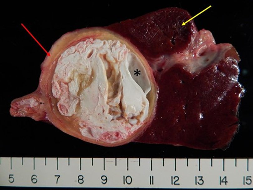
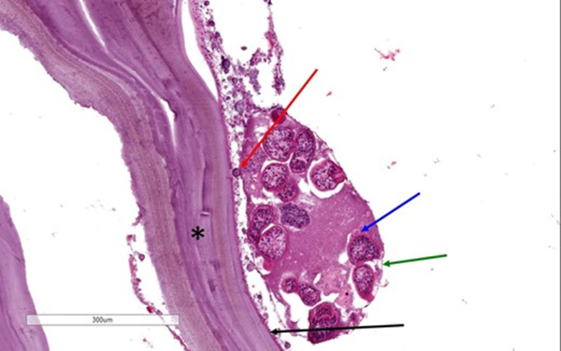
ECHINOCOCCAL CYST
The patient underwent total excision of the cyst followed by albendazole therapy for 4 weeks.
3/5
doi.org/10.1093/cid/ci…
#microbiology #GITwitter #MedStudentTwitter
The patient underwent total excision of the cyst followed by albendazole therapy for 4 weeks.
3/5
doi.org/10.1093/cid/ci…
#microbiology #GITwitter #MedStudentTwitter
A 29-YO♂️, 6 months before, HIV + & non–drug-resistant pulmonary tuberculosis, antiretroviral & 4-drug antituberculous therapy initiated but soon reduced to rifampin & isoniazid only: abdominal pain on the L side
CT: ?
1/5
DOI: 10.1056/NEJMicm2206174
#radiologist #GITwitter
CT: ?
1/5
DOI: 10.1056/NEJMicm2206174
#radiologist #GITwitter

CT: an enlarged spleen with numerous hypodense lesions (A).
CD4 cell count: 119/mm3
VIH viral load: 1778 copies/mm3
A splenectomy was performed to evaluate for cancer:
numerous necrotic nodules with purulent discharge (B).
2/5
#microbiology #gastroenterology #IDtwitter
CD4 cell count: 119/mm3
VIH viral load: 1778 copies/mm3
A splenectomy was performed to evaluate for cancer:
numerous necrotic nodules with purulent discharge (B).
2/5
#microbiology #gastroenterology #IDtwitter

🔬granulomatous inflammation with caseous necrosis (C) and acid-fast bacilli (D, arrowheads).
A tissue🧫: ➖
PCR: ➕for Mycobacterium tuberculosis
SPLENIC TUBERCULOSIS
3/5
#bacteriology #MedTwitter #GIPath
A tissue🧫: ➖
PCR: ➕for Mycobacterium tuberculosis
SPLENIC TUBERCULOSIS
3/5
#bacteriology #MedTwitter #GIPath

An 81-YO👵, type 2 DM: malaise, fever & anorexia, with oral pain & odynophagia, yellowish-white, pseudomembranous lesions on the tongue (A)
#endoscopy showed multiple shallow ulcers with a white coating (B)
1/6
doi.org/10.1503/cmaj.2…
#emergency #gastroenterology #MedEd
#endoscopy showed multiple shallow ulcers with a white coating (B)
1/6
doi.org/10.1503/cmaj.2…
#emergency #gastroenterology #MedEd

Glossal & esophageal🔬: multinucleated cells with moulded, ground-glass nuclei
PCR & immunohistochemical staining of the specimens: ➕ for herpes simplex virus type 1 (HSV-1)
HERPETIC GLOSSITIS AND ESOPHAGITIS
2/6
#GIPath #IDtwitter #microbiology
PCR & immunohistochemical staining of the specimens: ➕ for herpes simplex virus type 1 (HSV-1)
HERPETIC GLOSSITIS AND ESOPHAGITIS
2/6
#GIPath #IDtwitter #microbiology
Her hemoglobin A1c was 7.2%.
Doctors treated her with a 7-day course of acyclovir, intravenously because of her odynophagia, and the oral and esophageal lesions completely resolved.
3/6
#MedStudentTwitter #MedicalStudents
Doctors treated her with a 7-day course of acyclovir, intravenously because of her odynophagia, and the oral and esophageal lesions completely resolved.
3/6
#MedStudentTwitter #MedicalStudents
01/ Buckle up, everyone, it’s time for a Tweetorial. Been working on this one for a while. This time, I’ll focus on the most common mesenchymal malignancy of the digestive tract: gastrointestinal stromal tumor (GIST). #pathology #gipath #PathTwitter
02/ GIST arises from the interstitial cells of Cajal. It can originate anywhere in the GI tract, though most cases occur in the stomach or small bowel. Rectum cases are uncommon, and GIST is very rare in the esophagus, colon, or appendix.
03/ Old terms for GIST include GANT (gastrointestinal autonomic nerve tumor) and leiomyoblastoma. These terms are no longer used, though you may run across them in older literature.
@yaransarkis @MondayNightIBD @AmerGastroAssn @Spencerkelley7 @ayshaslam999 @jalpa_devi @purnie_mae @dunleavy_katie @KanikaGargMD @MarcelYibirin @JHaydek @DCharabaty @mjayoushe @AmCollegeGastro @ASGEendoscopy @Realcecum 8/ What’s in a C-scope ?
💎 #B2BPearl💎New guidelines @AmerGastroAssn
🔹Use High Def scope+++
🔹HDef scope ➕ Chromo (dye spray or virtual) if h/o dysplasia
🔹If using Standard Def: SD scope ➕Chromo dye spray only (not virtual)
🔗journals.lww.com/ajg/Abstract/2…
💎 #B2BPearl💎New guidelines @AmerGastroAssn
🔹Use High Def scope+++
🔹HDef scope ➕ Chromo (dye spray or virtual) if h/o dysplasia
🔹If using Standard Def: SD scope ➕Chromo dye spray only (not virtual)
🔗journals.lww.com/ajg/Abstract/2…
@yaransarkis @MondayNightIBD @AmerGastroAssn @Spencerkelley7 @ayshaslam999 @jalpa_devi @purnie_mae @dunleavy_katie @KanikaGargMD @MarcelYibirin @JHaydek @DCharabaty @mjayoushe @AmCollegeGastro @ASGEendoscopy @Realcecum 9/ What to biopsy ?
➕Targeted Bx🎯 of abnormal mucosa
➕Resection of polypoid lesion
➕Random Bx to document histologic extent/ healing
➕Extensive Random 4 quadrant bx every 10 cm IF no chromo, h/o dysplasia, poor visualization, PSC, foreshortened colon
➕Targeted Bx🎯 of abnormal mucosa
➕Resection of polypoid lesion
➕Random Bx to document histologic extent/ healing
➕Extensive Random 4 quadrant bx every 10 cm IF no chromo, h/o dysplasia, poor visualization, PSC, foreshortened colon
@yaransarkis @MondayNightIBD @AmerGastroAssn @Spencerkelley7 @ayshaslam999 @jalpa_devi @purnie_mae @dunleavy_katie @KanikaGargMD @MarcelYibirin @JHaydek @DCharabaty @mjayoushe @AmCollegeGastro @ASGEendoscopy @Realcecum 10/ What’s next after dx of Invisible dysplasia ?
💎 #B2BPearl💎
🔹Invisible dysplasia
→ Get 2nd opinion from expert #GIPath
→ HD CLN + dye chromo by expert endoscopist + Extensive non-targeted Bx if no resectable lesion seen
💎 #B2BPearl💎
🔹Invisible dysplasia
→ Get 2nd opinion from expert #GIPath
→ HD CLN + dye chromo by expert endoscopist + Extensive non-targeted Bx if no resectable lesion seen
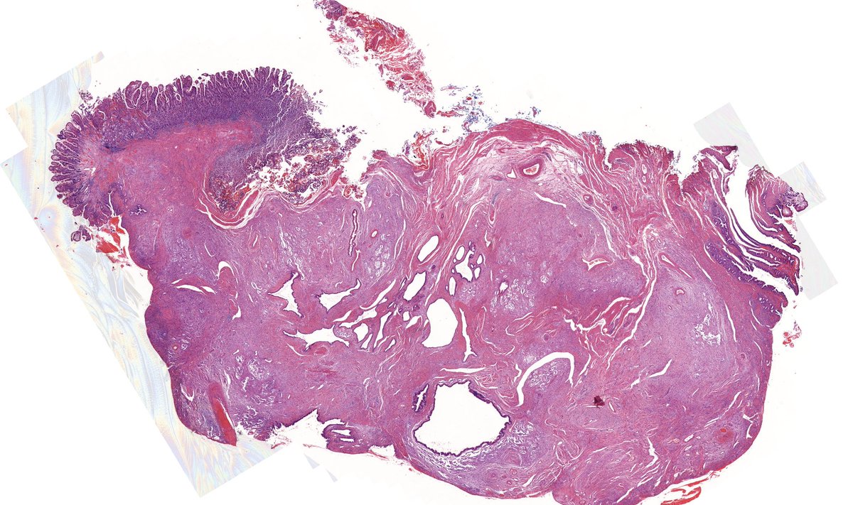
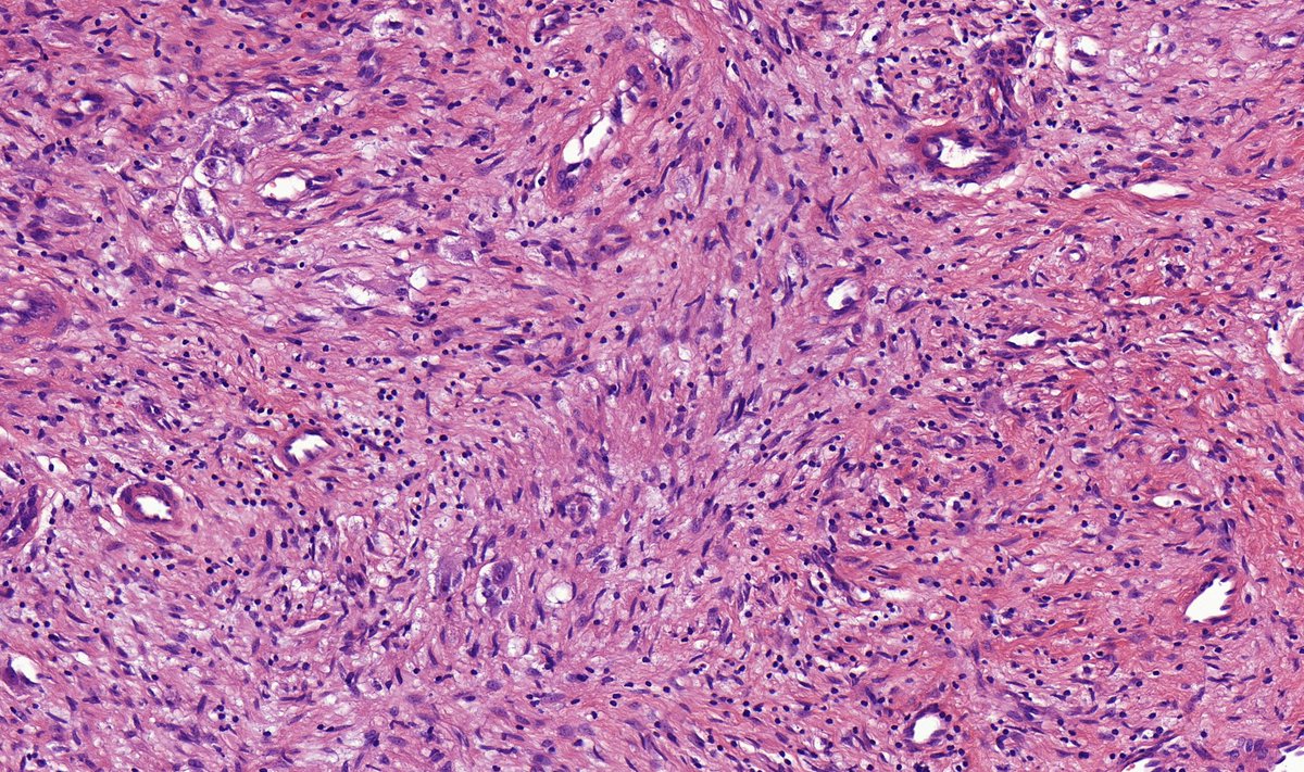
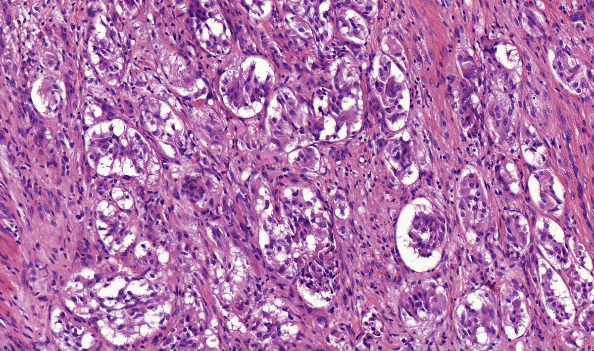
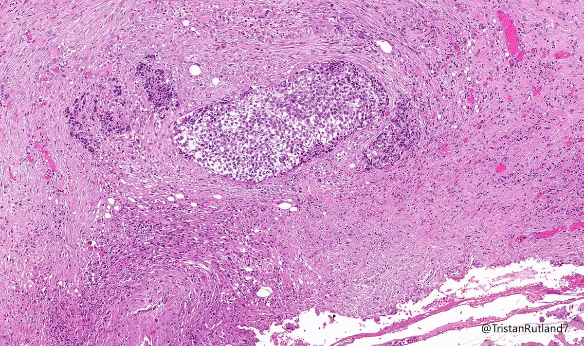
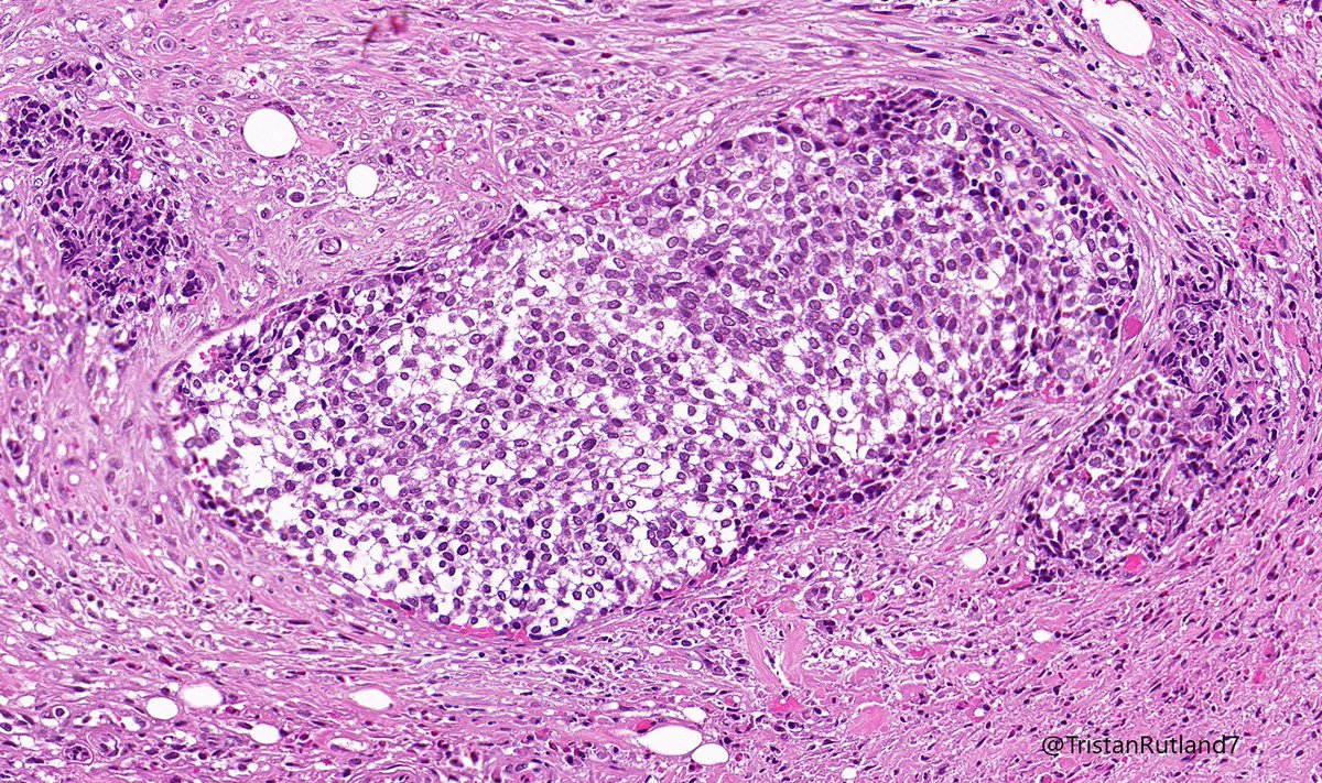
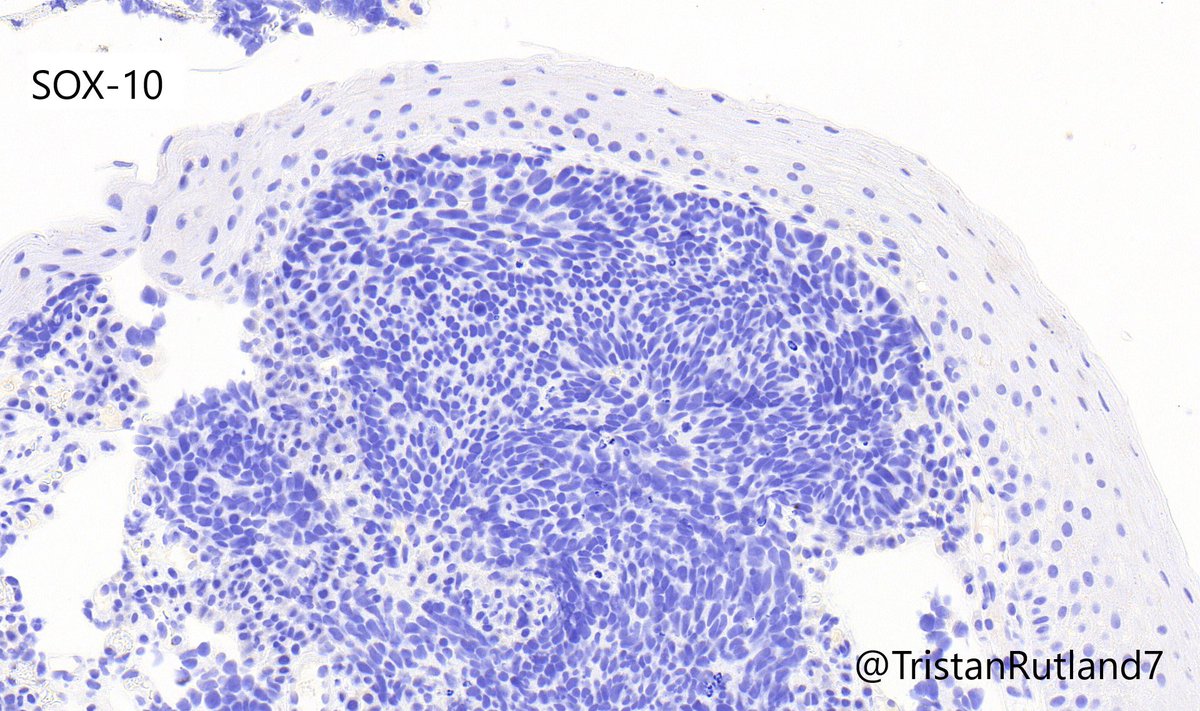
Question:
Based on the initial images, which of the following do you favour?
* Gastrointestinal stromal tumour
**Gangliocytic Paraganglioma
Based on the initial images, which of the following do you favour?
* Gastrointestinal stromal tumour
**Gangliocytic Paraganglioma
M 60ish. Caecal tumour ! Poorly differentiated , typical dMMR morphology ! MMR IHC - all four stains negative with adequate internal control ! How would you approach this? What would you do next in terms of work up or advice to clinicians ? #gipath #frcpath #PathTwitter 

Thank you all for engaging. This is really rare but once in a while, we do come across cases showing more than two markers' loss. Previously I had recommended genetic referral in such an instance thinking loss of MSH2/MSH6 is almost always germline i.e syndromic. 1/2
However, recently heard a talk on MSI & became aware that in most such cases, the MLH1 hypermethylation is a primary event and loss of MSH6 is secondary sporadic mutation. These patients in fact do not harbor germline mutation. Subsequently, I also found the following reference.
1/ Hello again, everyone. I have finally put together my third Tweetorial, and I will be focusing it on one single entity, the enigmatic and often frustrating Low-Grade Appendiceal Mucinous Neoplasm (LAMN). #pathology #GIpath 
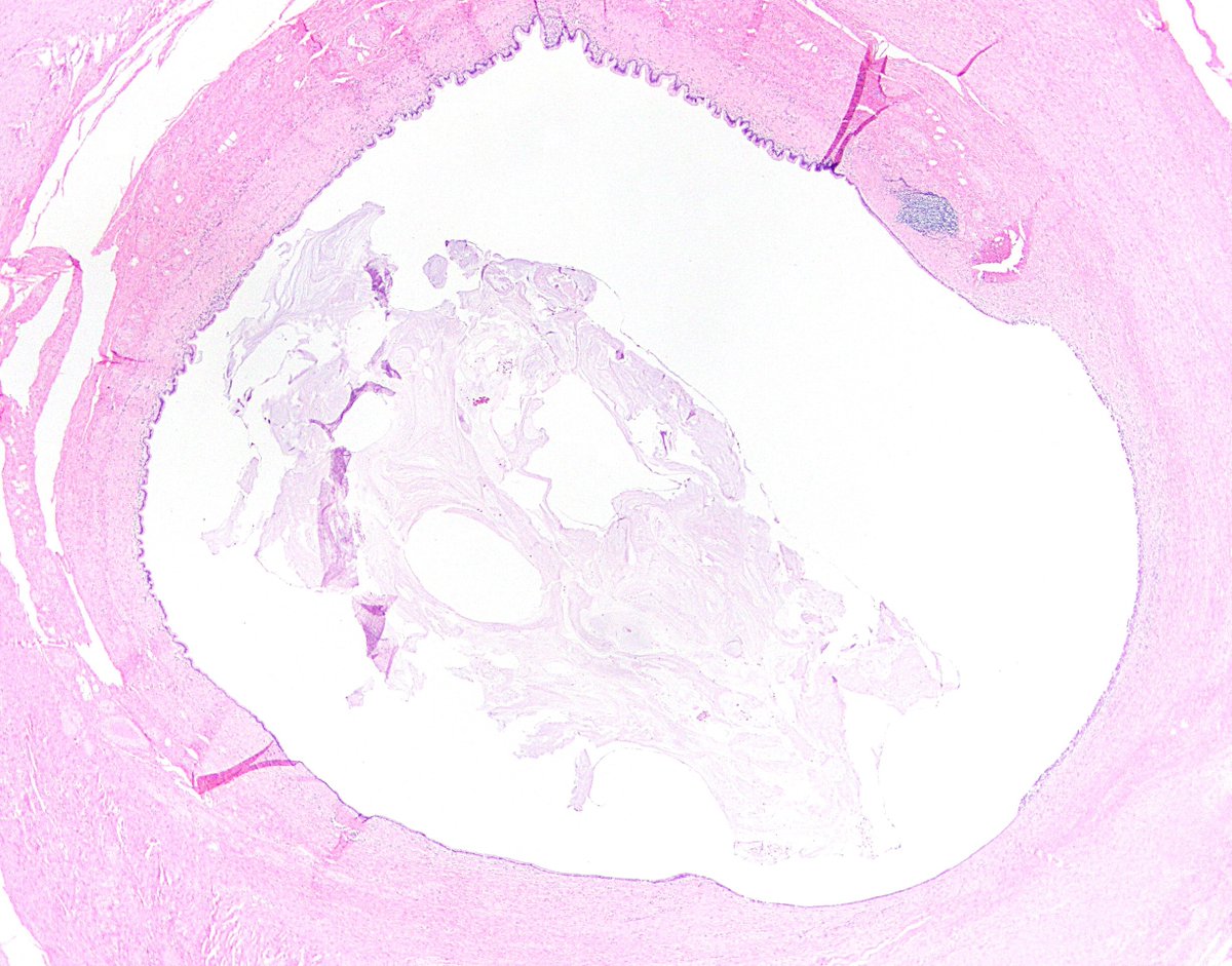
2/ For starters, what is a LAMN? The name gets you most of the way there. It technically is an appendiceal adenocarcinoma, because it is an epithelial malignancy composed of glandular epithelium.
3/ The key distinction is that LAMN invades by pushing rather than trickling/destructive invasion, so it’s not a low-grade “adenocarcinoma” like those we often see in the colorectum.
Question:
Based on the above image, what is your preferred diagnosis for this appendix?
*Low-grade appendiceal mucinous neoplasm
**High-grade appendiceal mucinous neoplasm
Based on the above image, what is your preferred diagnosis for this appendix?
*Low-grade appendiceal mucinous neoplasm
**High-grade appendiceal mucinous neoplasm
GOJ mass, older patient.
@ZHOUSEH @MattieFarzin @pembeoltulu @RunjanChetty @ariella8 @Dr_Brian_Cox @ALBoothMD @DrBMcGinn @DrGeeONE @MAHoureih @TheKarenPinto
#GIpath #PathTwitter #Surgpath


@ZHOUSEH @MattieFarzin @pembeoltulu @RunjanChetty @ariella8 @Dr_Brian_Cox @ALBoothMD @DrBMcGinn @DrGeeONE @MAHoureih @TheKarenPinto
#GIpath #PathTwitter #Surgpath


Further Hx for @Meghna0630
Male,
PMHx: skin cancer, DM type2, BPH
Presents with SBO. Nodule in stomach and small bowel (below).
Male,
PMHx: skin cancer, DM type2, BPH
Presents with SBO. Nodule in stomach and small bowel (below).

#gipath #granulomas #pathology
Ruptured crypt granuloma in UC.
1. LP - ruptured crypt; giant cell +
2. Deeper- histiocyte aggregate with only part of crypt seen (crypt loss)
@kriyer68 @smlungpathguy @pembeoltulu @RunjanChetty @ariella8 @DraEosina @DrMarkOng

Ruptured crypt granuloma in UC.
1. LP - ruptured crypt; giant cell +
2. Deeper- histiocyte aggregate with only part of crypt seen (crypt loss)
@kriyer68 @smlungpathguy @pembeoltulu @RunjanChetty @ariella8 @DraEosina @DrMarkOng
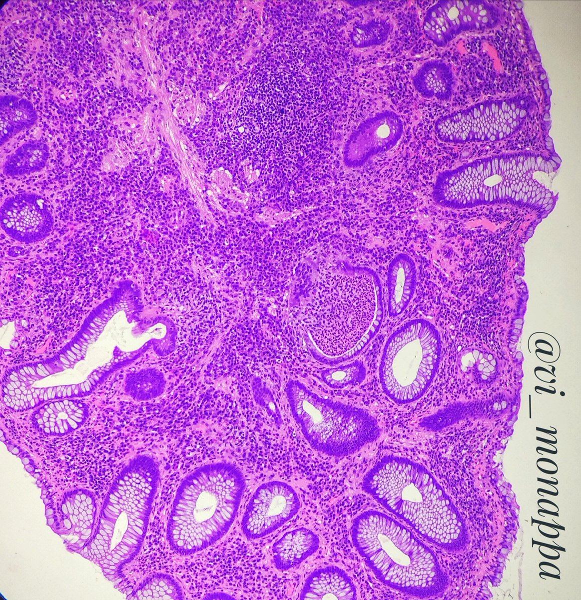
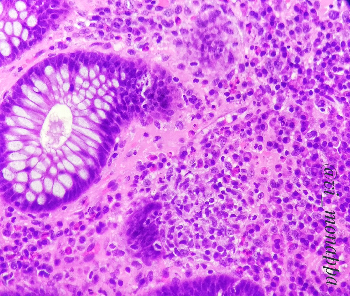
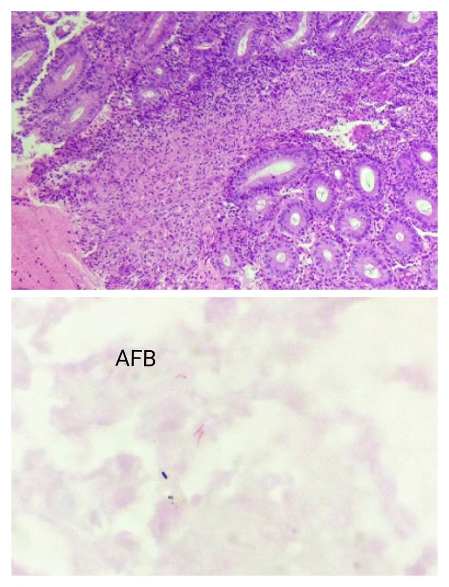
TB granulomas are large and confluent, may show necrosis.
AFB + confirms the diagnosis
@vhnguyenmd @anueru432 @anugnya_ran
AFB + confirms the diagnosis
@vhnguyenmd @anueru432 @anugnya_ran
@MondayNightIBD @SobiaMujtabaMD @DuekerJeffrey @DCharabaty 25y/oM quit🚬3 mos ago, now 3🩸loose BM/day,mild abdo cramps;Cousin w Crohns;Stool➖for infection;CLN: erythematous granular mucosa rectum+sigmoid, superficial ulcers;BX:Acute cryptitis,crypt abscess,crypt architecture distortion. What helps most dx UC vs Crohn’s?
#B2B #IBDPoll
#B2B #IBDPoll
@MondayNightIBD @SobiaMujtabaMD @DuekerJeffrey @DCharabaty UC and CD:
🔻Chronic inflammation of the GI tract
🔻Affects all ages: Typically starts between age 20-39
🔻Second peak of incidence age >50
🔻Flares of GI symptoms +/-systemic symptoms +/- EIM


🔻Chronic inflammation of the GI tract
🔻Affects all ages: Typically starts between age 20-39
🔻Second peak of incidence age >50
🔻Flares of GI symptoms +/-systemic symptoms +/- EIM
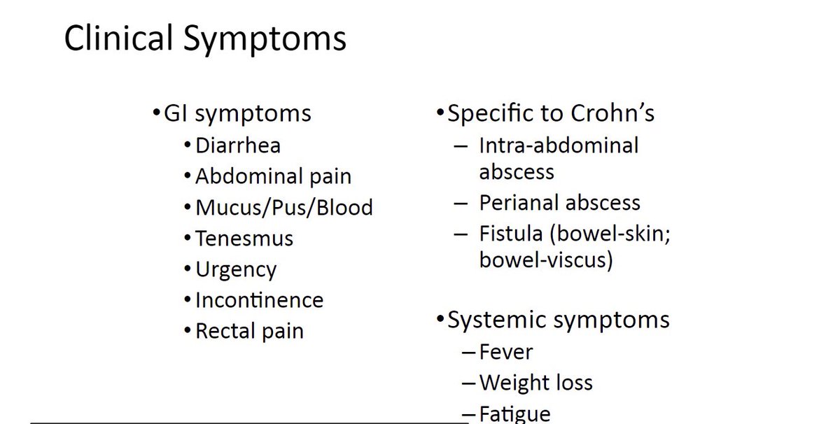
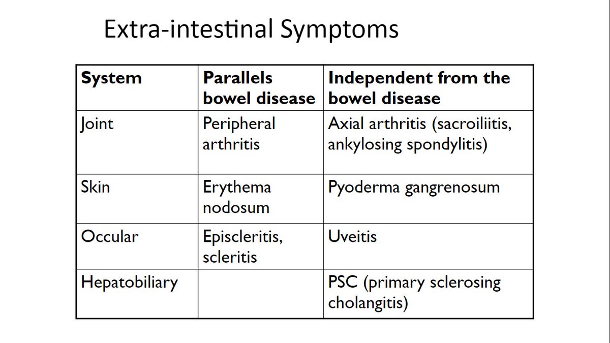
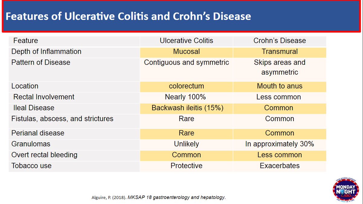
@MondayNightIBD @SobiaMujtabaMD @DuekerJeffrey @DCharabaty 3/ CD:
💡Skipped lesion, any part of GI tract
💡Most common:Colon+ileum
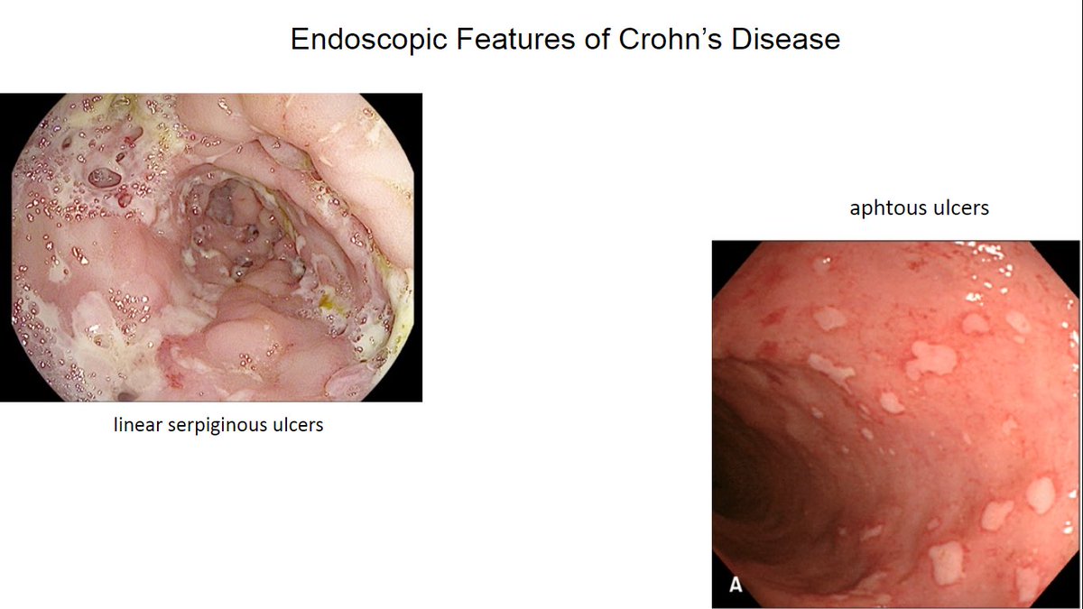
Hallmark➡️ulcers: aphthous,deep large/linear/serpiginous
💡Transmural inflamm -> stricturing, perforating dis.
🚩#B2BPearl
👉🏼Rectum can be involved in CD;➕anorectal ulcers → ⬆️risk of perianal disease

💡Skipped lesion, any part of GI tract
💡Most common:Colon+ileum
Hallmark➡️ulcers: aphthous,deep large/linear/serpiginous
💡Transmural inflamm -> stricturing, perforating dis.
🚩#B2BPearl
👉🏼Rectum can be involved in CD;➕anorectal ulcers → ⬆️risk of perianal disease
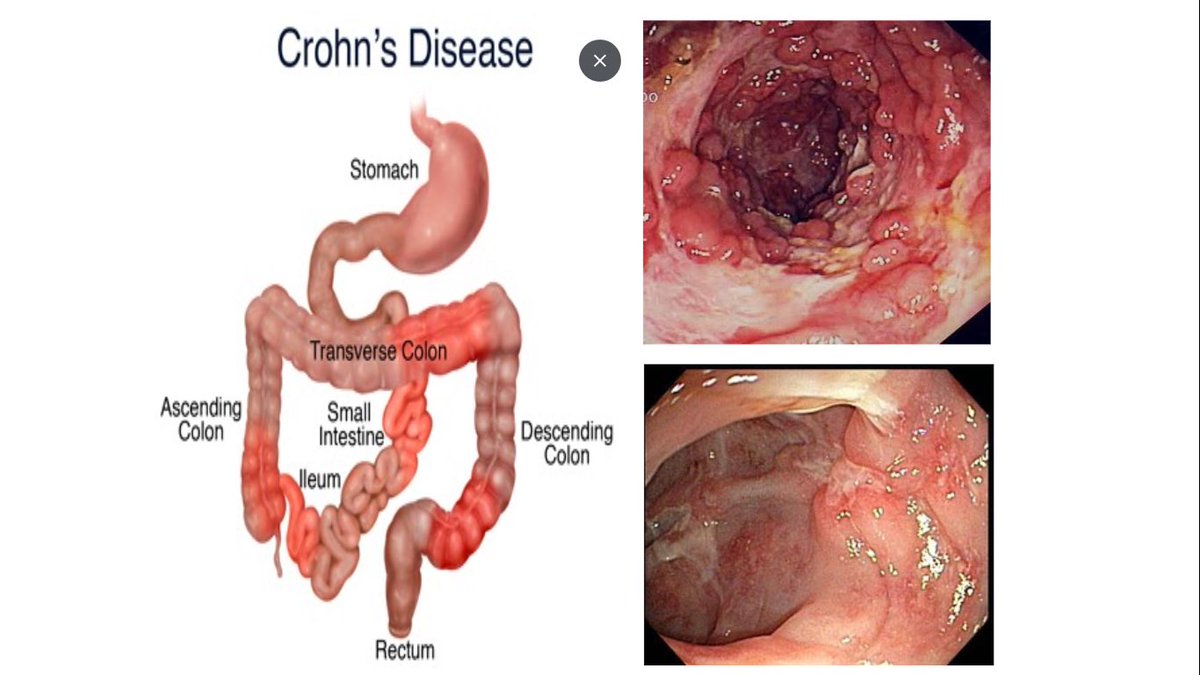
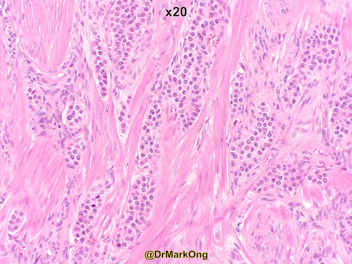
How best to classify this gastric tumour? #GIpath
>95% of tumour is a WD NET (like picture 1), <5% shows mucin & goblet cells (pictures 2-4). Complicated by neoadjuvant chemotherapy for adenocarcinoma (presumably index biopsies looked more epithelial). IHC next tweet...


>95% of tumour is a WD NET (like picture 1), <5% shows mucin & goblet cells (pictures 2-4). Complicated by neoadjuvant chemotherapy for adenocarcinoma (presumably index biopsies looked more epithelial). IHC next tweet...
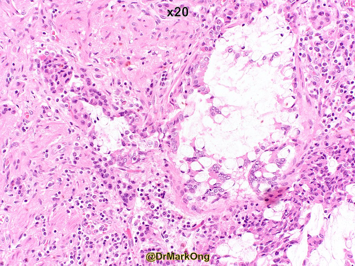
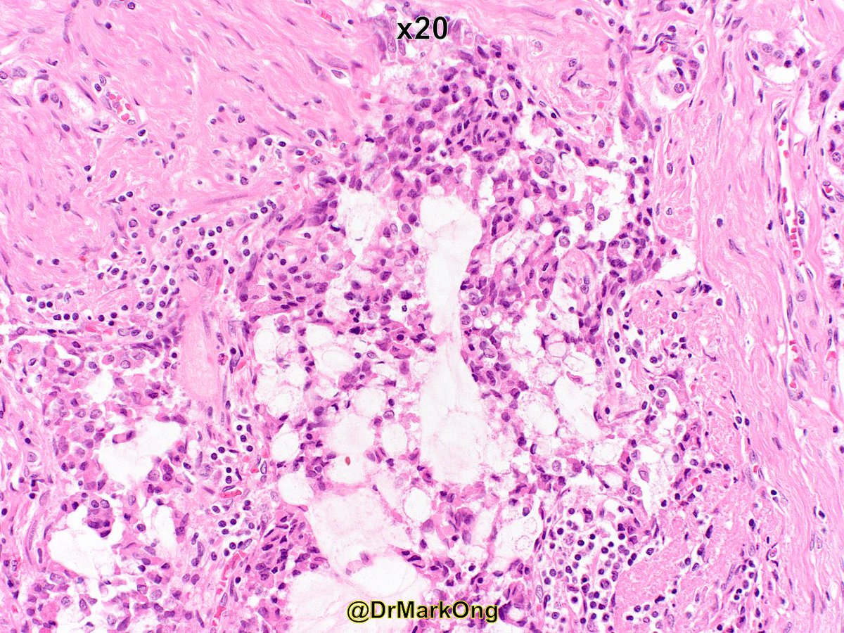
Synaptophysin -ve in epithelial component. Chromogranin strong +ve in NE component, weak in epithelial component. Ki67 high in epithelial component, low in NE component.
Are the mucin-producing areas amphicrine (dual differentiation in 1 cell) or a separate epithelial component?


Are the mucin-producing areas amphicrine (dual differentiation in 1 cell) or a separate epithelial component?


Definition of mixed neuroendocrine, non-neuroendocrine neoplasm:
⏺️2 discrete components (morphologically & immunohistochemically)
⏺️Each component >30% (small cell carcinoma is an exception)
⏺️2 discrete components (morphologically & immunohistochemically)
⏺️Each component >30% (small cell carcinoma is an exception)
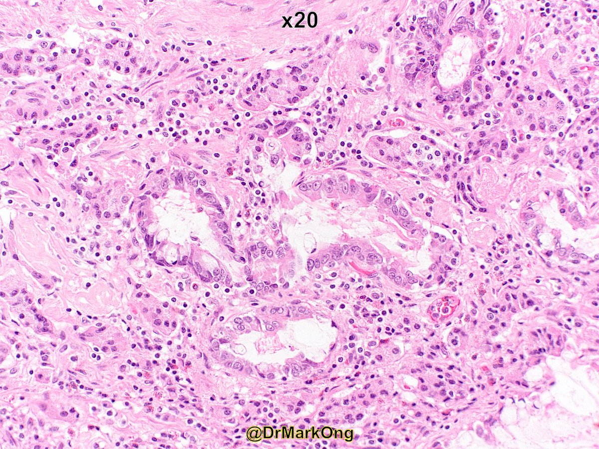
Any guesses? Site? Diagnosis?#SurgPath #GIpath #PathTwitter @Teclis82 @pezhouh @CArnold_GI @mhassanaimc @ac_pathgal @ALBoothMD @SwikrityUMD @D_KumarMD @Gagandeepk5MD 

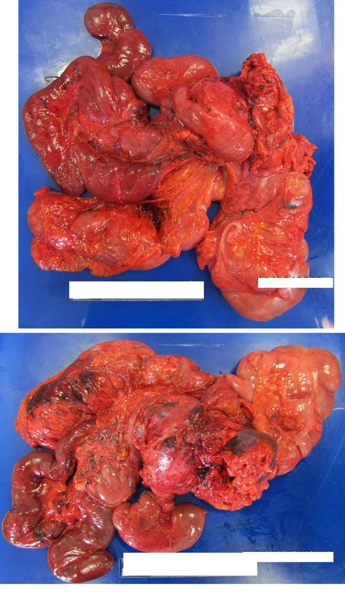
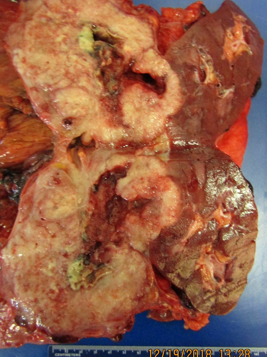
Resident Midnight Horror:
My colleague (PGY-1) received this specimen while on call:
A whipple specimen resected with roux-en-Y procedure, right nephrectomy, and right hemicolectomy, all attached together!!
Try orient the specimen. #grosspath #GIpath #PathTwitter #SurgPath

My colleague (PGY-1) received this specimen while on call:
A whipple specimen resected with roux-en-Y procedure, right nephrectomy, and right hemicolectomy, all attached together!!
Try orient the specimen. #grosspath #GIpath #PathTwitter #SurgPath
Young lady with weakness and anemia.
Endoscopy – diffuse scalloping of the duodenal mucosa
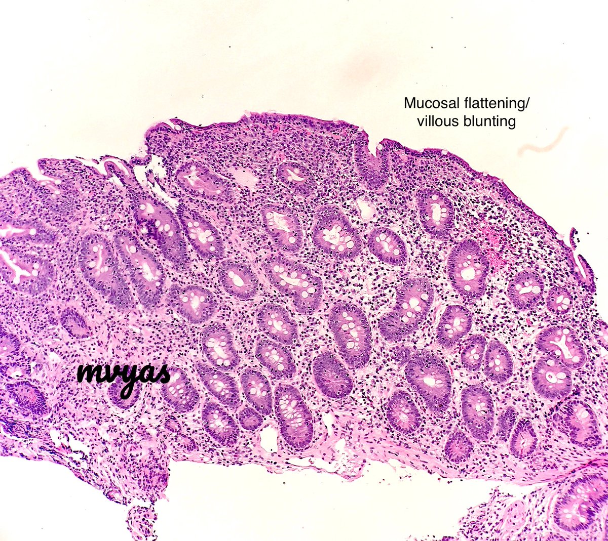
Biopsy – Mucosal flattening
Crypt hyperplasia
Intraepithelial lymphocytosis.
IgA anti-tissue transglutaminase (anti-TTG) NEGATIVE.
So, is this not celiac disease (CD)?
#GIpath #pathboards


Endoscopy – diffuse scalloping of the duodenal mucosa
Biopsy – Mucosal flattening
Crypt hyperplasia
Intraepithelial lymphocytosis.
IgA anti-tissue transglutaminase (anti-TTG) NEGATIVE.
So, is this not celiac disease (CD)?
#GIpath #pathboards



High clinical suspicion for CD -check IgA levels
In IgA deficiency, IgA-TTG and IgA-EMA (anti-endomysial Ab) will be negative
Alternative tests - IgG-DGP (deaminated gliadin peptide)/ IgG TTG
HLA-DQ typing - HLA-DQ2 (~95% pts) and HLA-DQ8
#GIpath #pathboards
In IgA deficiency, IgA-TTG and IgA-EMA (anti-endomysial Ab) will be negative
Alternative tests - IgG-DGP (deaminated gliadin peptide)/ IgG TTG
HLA-DQ typing - HLA-DQ2 (~95% pts) and HLA-DQ8
#GIpath #pathboards
Lastly, here's the colon bx of this pt showing lymphocytic colitis pattern
Microscopic colitis is more prevalent in pts w/ CD
Pts w/ MC and CD often have more severe histologic features, may require steroid rx
Response of MC to gluten free diet is not known.
#GIpath #pathboards
Microscopic colitis is more prevalent in pts w/ CD
Pts w/ MC and CD often have more severe histologic features, may require steroid rx
Response of MC to gluten free diet is not known.
#GIpath #pathboards

🤩I am constantly marveled by the endless ways in which we #Pathology and #LabMedicine, can use Twitter to engage, share, support & learn from each other. Here is the link to my presentation that celebrates the unlimited opportunities to harness Twitter👉🏽
bit.ly/39kA627
bit.ly/39kA627

Like the #SolarEclipse that occurred in August 2017, to me, Twitter has been a community wide experience of marveling a visual process that can create a burst of awe at an organic velocity. It's really exciting to be engaged in this global experience. 

#AcademicTwitter is using Twitter at the University and Research setting to teach. It has wide range benefits and I recommend reading the following article written by @soragnilab and @Aiims1742 published in @nature that describes this phenomenon. doi.org/10.1038/s41568… 



1/
Necrosis = cell death (unlike apoptosis, it does not occur naturally and is not programmed)
This short #Tweetorial shows you some of the histologic flavors of necrosis. The stain in each of these pics is hematoxylin-eosin (H&E)
#pathology #pulmpath #pathtweetorial
Necrosis = cell death (unlike apoptosis, it does not occur naturally and is not programmed)
This short #Tweetorial shows you some of the histologic flavors of necrosis. The stain in each of these pics is hematoxylin-eosin (H&E)
#pathology #pulmpath #pathtweetorial
2/
First a question to test your knowledge. Necrosis with large numbers of neutrophils is called:
First a question to test your knowledge. Necrosis with large numbers of neutrophils is called:


