Discover and read the best of Twitter Threads about #RadTwitter
Most recents (24)
Up for a Sunday tweetorial? 🤓
If you see this multicystic lung lesion 🫁 in the posterobasal region of the lower lobes in a young patient 🚹🚺with no pathological history, two differential diagnoses should be considered: CPAM vs pulmonary sequestration ✅
#radiology #FOAMrad
If you see this multicystic lung lesion 🫁 in the posterobasal region of the lower lobes in a young patient 🚹🚺with no pathological history, two differential diagnoses should be considered: CPAM vs pulmonary sequestration ✅
#radiology #FOAMrad

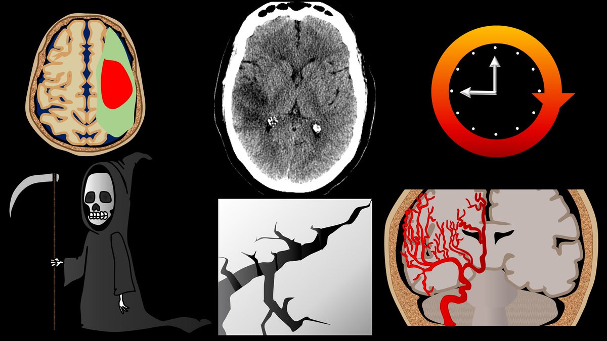
1/Time is brain! So you don’t have time to struggle w/that stroke alert head CT.
Here’s a #tweetorial to help you with the CT findings in acute stroke.
#medtwitter #FOAMed #FOAMrad #ESOC #medstudent #neurorad #radres #meded #radtwitter #stroke #neurology #neurotwitter
Here’s a #tweetorial to help you with the CT findings in acute stroke.
#medtwitter #FOAMed #FOAMrad #ESOC #medstudent #neurorad #radres #meded #radtwitter #stroke #neurology #neurotwitter
1/Don’t let all your effort be in VEIN!
Developmental venous anomalies (DVAs) are often thought incidental but ignore them at your own risk!
A #tweetorial about how to know when DVAs are the most important finding
#meded #medtwitter #neurorad #neurotwitter #radtwitter #radres
Developmental venous anomalies (DVAs) are often thought incidental but ignore them at your own risk!
A #tweetorial about how to know when DVAs are the most important finding
#meded #medtwitter #neurorad #neurotwitter #radtwitter #radres

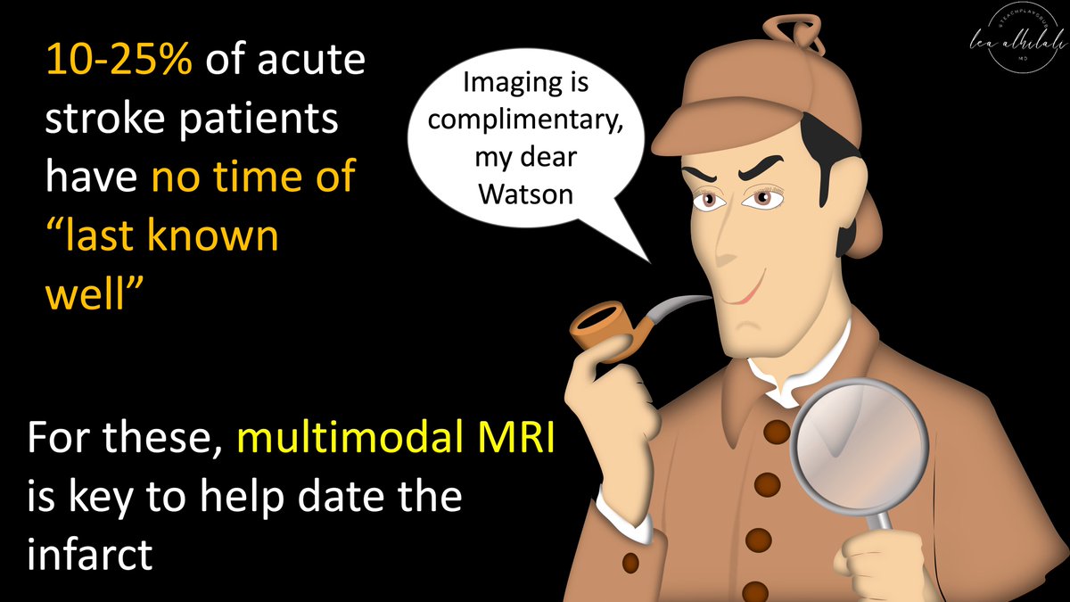
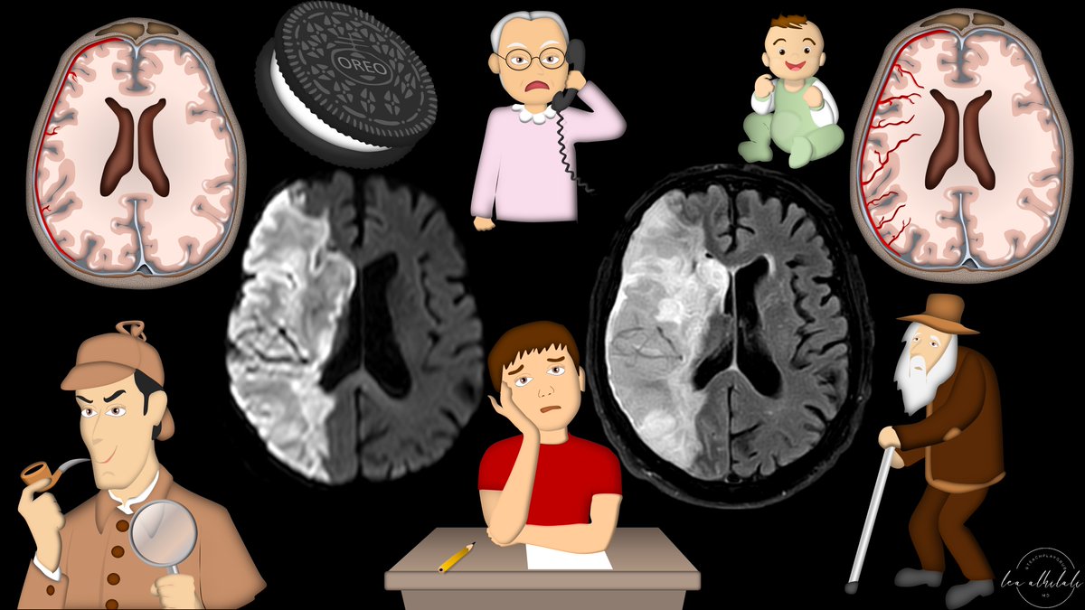
1/Time is brain! But what time is it?
If you don’t know the time of stroke onset, are you able to deduce it from imaging?
Here’s a #tweetorial to help you date a #stroke on MR!
#medtwitter #meded #neurotwitter #neurology #neurorad #radres #radtwitter #radiology #FOAMed #FOAMrad
If you don’t know the time of stroke onset, are you able to deduce it from imaging?
Here’s a #tweetorial to help you date a #stroke on MR!
#medtwitter #meded #neurotwitter #neurology #neurorad #radres #radtwitter #radiology #FOAMed #FOAMrad
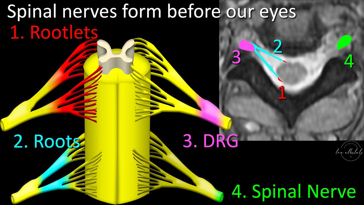
1/Feeling unarmed when it comes to evaluating cervical radiculopathy & foraminal narrowing on MR?
Here’s a #tweetorial that’ll take that weight off your shoulder & show you how to rate cervical foraminal stenosis!
#medtwitter #meded #FOAMed #radtwitter #neurorad #spine #radres
Here’s a #tweetorial that’ll take that weight off your shoulder & show you how to rate cervical foraminal stenosis!
#medtwitter #meded #FOAMed #radtwitter #neurorad #spine #radres

1/I call the skullbase “homebase” bc you can’t make an anatomy homerun without it!
Most know the arteries of the skullbase, but few know the veins. Do you?
Here’s a🧵to help you remember #skullbase venous #anatomy!
#medtwitter #meded #neurorad #radtwitter #neurosurgery #radres
Most know the arteries of the skullbase, but few know the veins. Do you?
Here’s a🧵to help you remember #skullbase venous #anatomy!
#medtwitter #meded #neurorad #radtwitter #neurosurgery #radres

1/Does trying to remember inferior frontal gyrus anatomy leave you speechless?
Do you get a Broca’s aphasia trying to name the parts?
Here’s a #tweetorial to help you remember the #anatomy of this important region
#medtwitter #meded #neurotwitter #neurorad #radtwitter #radres
Do you get a Broca’s aphasia trying to name the parts?
Here’s a #tweetorial to help you remember the #anatomy of this important region
#medtwitter #meded #neurotwitter #neurorad #radtwitter #radres

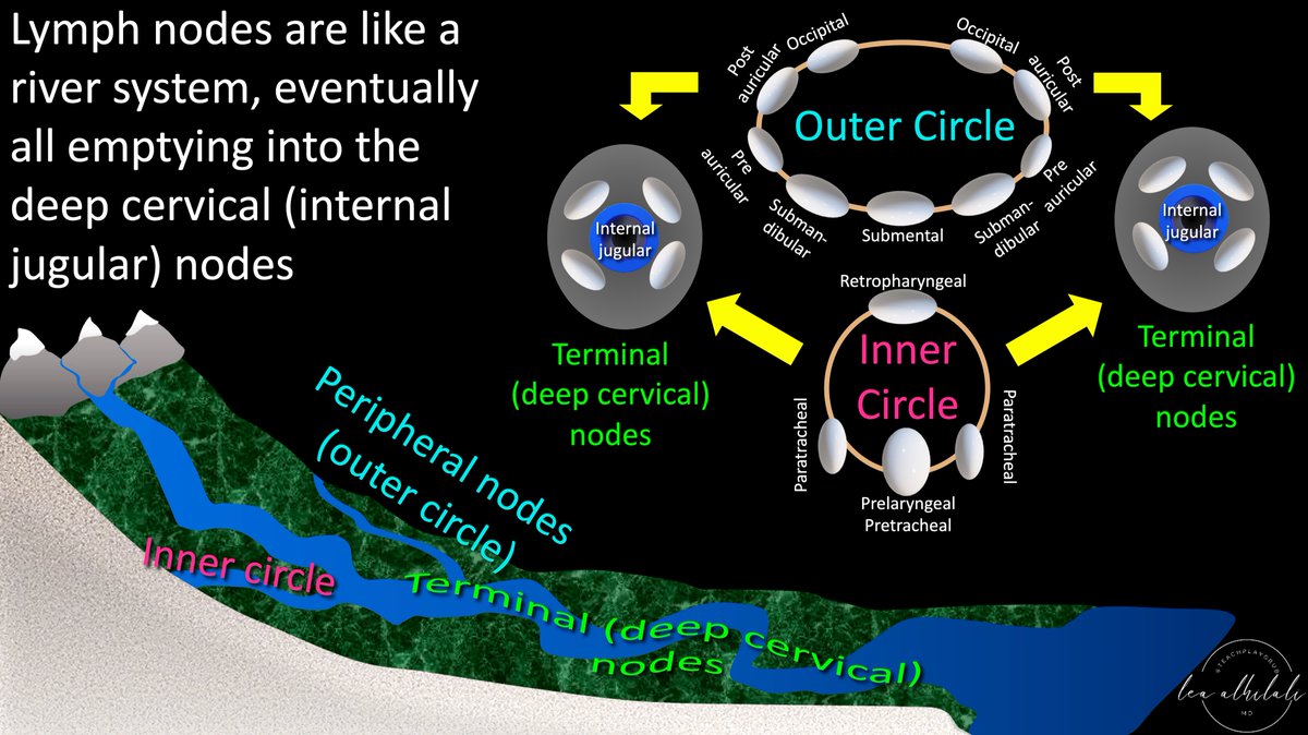
1/To be or not 2b?? That is the question!
Do you have questions about how to remember cervical lymph node anatomy & levels?
Here’s a #tweetorial to show you how--#Superbowl weekend edition!
#medtwitter #meded #neurorad #HNrad #FOAMed #FOAMrad #radres #radtwitter #ENT #radiology
Do you have questions about how to remember cervical lymph node anatomy & levels?
Here’s a #tweetorial to show you how--#Superbowl weekend edition!
#medtwitter #meded #neurorad #HNrad #FOAMed #FOAMrad #radres #radtwitter #ENT #radiology

1/If all you know is: To Zanzibar By Motor Car—then you don’t even know half of facial nerve anatomy—literally!
Here’s a #tweetorial on the facial nerve anatomy you don’t know!
#medtwitter #neurotwitter #neurorad #radres #meded #FOAMed #neurosurgery #neurology #radtwitter
Here’s a #tweetorial on the facial nerve anatomy you don’t know!
#medtwitter #neurotwitter #neurorad #radres #meded #FOAMed #neurosurgery #neurology #radtwitter

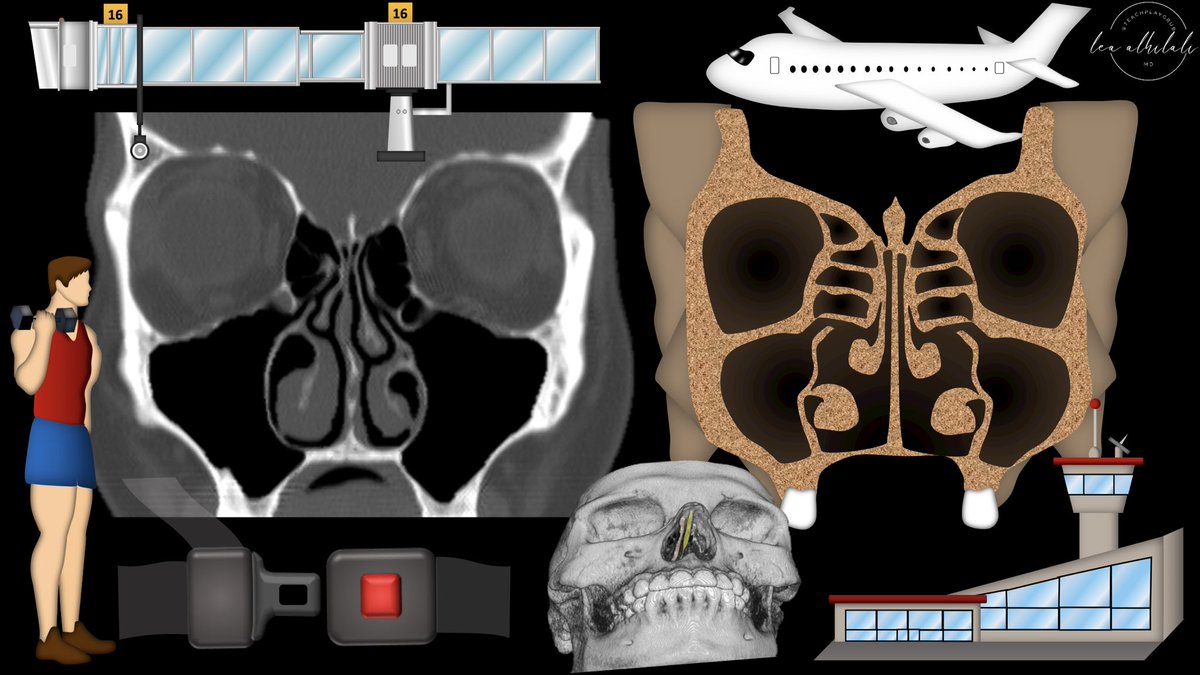
1/Time to FESS up! Do you understand functional endoscopic sinus surgery (FESS)?
If you read sinus CTs, you must know what they’re doing to make the helpful findings
Here’s a #tweetorial to help you!
#medtwitter #meded #FOAMed #FOAMrad #radres #neurorad #HNrad #radtwitter
If you read sinus CTs, you must know what they’re doing to make the helpful findings
Here’s a #tweetorial to help you!
#medtwitter #meded #FOAMed #FOAMrad #radres #neurorad #HNrad #radtwitter
31yo F, brother sudden cardiac death at 34yo, father sudden cardiac death at 45yo
Should she be worried??
#radtwitter #medtwitter #cardiotwitter #whyCMR
Should she be worried??
#radtwitter #medtwitter #cardiotwitter #whyCMR
Patient also had basal septal hypertrophy up to 21mm (left), associated with delayed enhancement (right) 


Dad was thought to have died from MI. Brother had suspicion of ARVD on echo, died before CMR, no autopsy. Her CMR was requested to rule out ARVD.
21 month old male, cough, prolonged fever, normal chest X-ray, abnormal echo
#radtwitter #medtwitter #pedsrad #cardiotwitter
#radtwitter #medtwitter #pedsrad #cardiotwitter
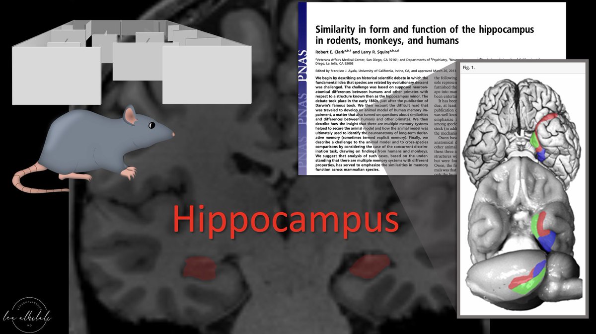
1/Is your ability to remember temporal lobe anatomy seem, well, temporary?
Here’s a #tweetorial to help you remember the structures of the temporal lobe!
#medtwitter #meded #neurotwitter #radtwitter #radres #neurorad #FOAMed #neurosurgery #medstudenttwitter #neurology
Here’s a #tweetorial to help you remember the structures of the temporal lobe!
#medtwitter #meded #neurotwitter #radtwitter #radres #neurorad #FOAMed #neurosurgery #medstudenttwitter #neurology

1/Nothing strikes fear into the heart of a radiologist like the question,“Is it safe to do an MRI on this pt w/an implanted device?”
Never fear again! Here’s a #tweetorial on how to navigate implanted devices & #MRI
#medtwitter #meded #radtwitter #radres #neurotwitter #neurorad
Never fear again! Here’s a #tweetorial on how to navigate implanted devices & #MRI
#medtwitter #meded #radtwitter #radres #neurotwitter #neurorad

62 y/o M presents with signs of raised intracranial pressure. CT shows a hyper dense mass crossing the corpus callosum. On MR, the mass is hypointense on T2WI, restricting diffusion, and homogenously enhancing along the periventricular surface.
#radtwitter #radres #neurotwitter


#radtwitter #radres #neurotwitter


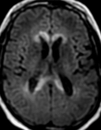

The corpus callosum is composed of very dense white matter tracks. Only aggressive tumors or lesions that effect the white matter cross the midline through the CC.
Diff Diagnosis for CC masses
High grade astrocytoma/Glioblastoma
Primary CNS lymphoma
Tumefactive Demyelination
Diff Diagnosis for CC masses
High grade astrocytoma/Glioblastoma
Primary CNS lymphoma
Tumefactive Demyelination
Dx: Primary CNS lymphoma
PCNSL has a highly variable imaging appearance. Classically, it presents as a hyperdense mass with restricted diffusion and relatively hypointensity on T2WI due to hypercellularity. The mass enhances homogeneously and may cross the CC. #Neurosurgery
PCNSL has a highly variable imaging appearance. Classically, it presents as a hyperdense mass with restricted diffusion and relatively hypointensity on T2WI due to hypercellularity. The mass enhances homogeneously and may cross the CC. #Neurosurgery
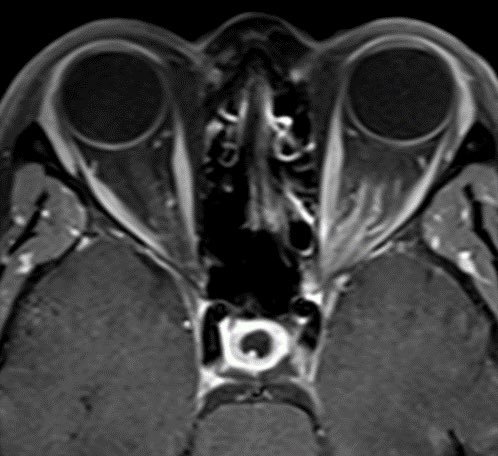
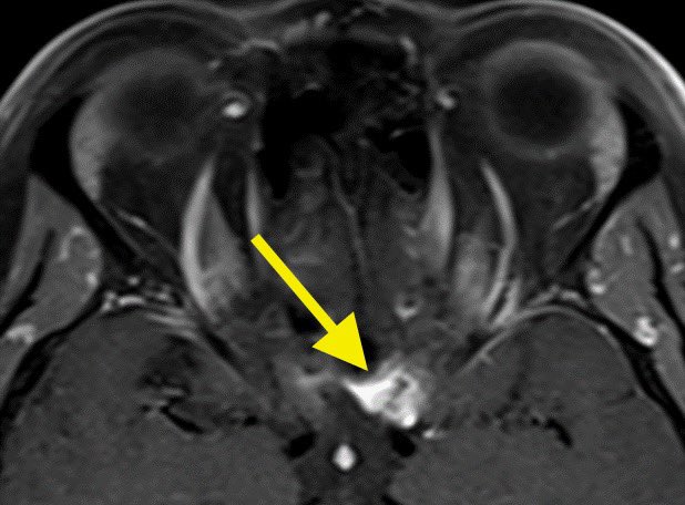
45F presents with painless progressive left eye vision loss. MR shows homogenous enhancement encasing the left optic nerve with an associated lesion at the entrance of the optic canal (yellow arrow)
#radres #futureradres #NeuroRad #MedTwitter @AlbanyMedRadRes


#radres #futureradres #NeuroRad #MedTwitter @AlbanyMedRadRes

Differential Diagnosis:
Optic Neuritis
Optic nerve sheath meningioma
Optic nerve glioma
Orbital sarcoidosis
Orbital lymphoma
Orbital pseudotumor
#Ophthalmology #radtwitter
Optic Neuritis
Optic nerve sheath meningioma
Optic nerve glioma
Orbital sarcoidosis
Orbital lymphoma
Orbital pseudotumor
#Ophthalmology #radtwitter
Diagnosis: Optic nerve sheath meningioma
Remember the optic nerve is an extension of the CNS and therefore, is surrounded by meninges and arachnoid cap cells from which meningiomas arise. Look for the “tram-track” sign of enhancement surrounding the optic nerve
#Ophthalmology
Remember the optic nerve is an extension of the CNS and therefore, is surrounded by meninges and arachnoid cap cells from which meningiomas arise. Look for the “tram-track” sign of enhancement surrounding the optic nerve
#Ophthalmology
23 yr old with headache. MR shows a “bubbly” mass in the right lateral ventricle near the foramen of Monro. The mass abuts the septum pellucidum and displays mild contrast enhancement.
#neurotwitter #radtwitter #RadEd #MedTwitter #radres @TheASNR @ESNRad @ASHNRSociety


#neurotwitter #radtwitter #RadEd #MedTwitter #radres @TheASNR @ESNRad @ASHNRSociety


Differential diagnosis:
Subependymoma
Choroid plexus neoplasm
Central neurocytoma
Intraventricular meningioma
Mets
Subependymoma
Choroid plexus neoplasm
Central neurocytoma
Intraventricular meningioma
Mets
Answer: confirmed central neurocytoma
Classically, look for the “bubbly” mass abutting/attached to the septum pellucidum near the foramen of monro with enhancement.
#futureradres
Classically, look for the “bubbly” mass abutting/attached to the septum pellucidum near the foramen of monro with enhancement.
#futureradres
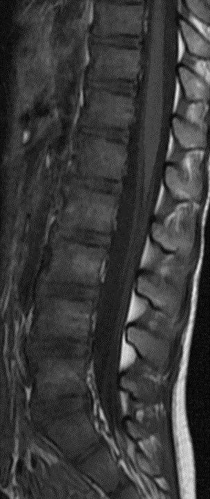
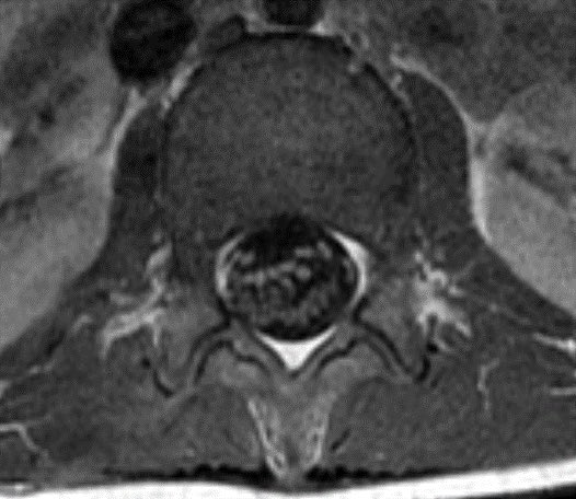
Child presents with weakness. MR shows enhancement of the pial surface of the conus and ventral cauda equina nerve roots.
#radtwitter #MedTwitter #radres #futureradres #Pediatrics #Neurology @TheASNR @The_ASPNR @AlbanyMedRadRes


#radtwitter #MedTwitter #radres #futureradres #Pediatrics #Neurology @TheASNR @The_ASPNR @AlbanyMedRadRes

Differential diagnosis:
Leptomeningeal carcinomatosis
Lymphoma
Arachnoiditis (all causes)
Guillain-barre
Neurosarcoidosis
Leptomeningeal carcinomatosis
Lymphoma
Arachnoiditis (all causes)
Guillain-barre
Neurosarcoidosis
Diagnosis: Guillain-Barré syndrome
These are the typical imaging features for GBS. Contrast is absolutely necessary.
There was no history to suggest systemic sarcoidosis, malignancy, or recent procedure (risk factor for spinal meningitis/arachnoiditis)
These are the typical imaging features for GBS. Contrast is absolutely necessary.
There was no history to suggest systemic sarcoidosis, malignancy, or recent procedure (risk factor for spinal meningitis/arachnoiditis)
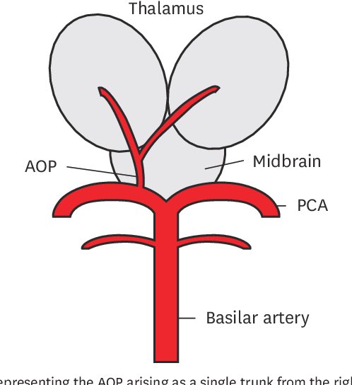
Patient presents with altered mental status. Unenhanced CT shows discrete hypodense foci in the bilateral paramedian thalami
#radres #futureradres #radtwitter @ACRRFS @RSNA
#radres #futureradres #radtwitter @ACRRFS @RSNA

Differential diagnosis includes:
Top of the basilar artery syndrome
Artery of Percheron infarct
Bilateral internal cerebral vein thrombosis
Top of the basilar artery syndrome
Artery of Percheron infarct
Bilateral internal cerebral vein thrombosis
80 yo ♀️ with chronic cough.
What would you do with this "ugly" lung lesion? 🧵👇🏻
#radres #radtwitter #radiology #chestrad
What would you do with this "ugly" lung lesion? 🧵👇🏻
#radres #radtwitter #radiology #chestrad

❓❓❓
1/My hardest #tweetorial yet! Are you up for the challenge?
How stroke perfusion imaging works!
Ever wonder why it’s Tmax & not Tmin? Do you not question & let RAPID read the perfusion for you? Not anymore!
#stroke #neurotwitter #neurorad #meded #FOAMed #radtwitter #medtwitter
How stroke perfusion imaging works!
Ever wonder why it’s Tmax & not Tmin? Do you not question & let RAPID read the perfusion for you? Not anymore!
#stroke #neurotwitter #neurorad #meded #FOAMed #radtwitter #medtwitter

55 yr old with stuffy nose. CT demonstrates a destructive mass centered in the olfactory recess with extension through the cribriform plate. MR shows avid enhancement of the invasive mass.
#radres #NeuroRad #radtwitter #MedTwitter


#radres #NeuroRad #radtwitter #MedTwitter


Differential:
Sinonasal carcinoma (SCC, SNUC, adenocarcinoma)
Esthesioneuroblastoma
Lymphoma
Mets
Sinonasal carcinoma (SCC, SNUC, adenocarcinoma)
Esthesioneuroblastoma
Lymphoma
Mets
Answer: confirmed Esthesioneuroblastoma
Esthesioneuroblastoma presents as a destructive mass with intracranial extension and waist centered at the cribriform plate. Peritumoral cysts at the tumor-brain interface are classic though were not seen in this case.
Esthesioneuroblastoma presents as a destructive mass with intracranial extension and waist centered at the cribriform plate. Peritumoral cysts at the tumor-brain interface are classic though were not seen in this case.
1/”I LOVE spinal cord syndromes!” is a phrase that has NEVER, EVER been said by anyone.
Never fear—here is a #tweetorial on all the incomplete #spinalcord syndromes!
#medtwitter #neurotwitter #neurology #neurosurgery #neurorad #radres #meded #FOAMed #FOAMrad #radtwitter #spine
Never fear—here is a #tweetorial on all the incomplete #spinalcord syndromes!
#medtwitter #neurotwitter #neurology #neurosurgery #neurorad #radres #meded #FOAMed #FOAMrad #radtwitter #spine

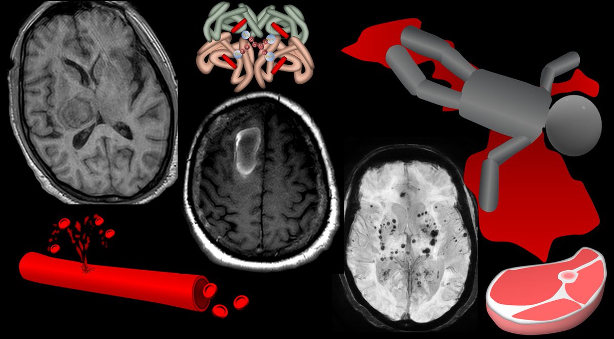
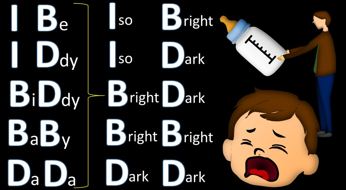
1/Asking “How old are you” can be dicey—both in real life & on MRI! Do you know how to tell the age of blood on MRI?
Here’s a #tweetorial on how to date blood on MRI
#medtwitter #neurorad #radtwitter #RSNA2022 #RSNA22 #radres #neurosurgery #neurology #meded #neurotwitter #FOAMed
Here’s a #tweetorial on how to date blood on MRI
#medtwitter #neurorad #radtwitter #RSNA2022 #RSNA22 #radres #neurosurgery #neurology #meded #neurotwitter #FOAMed
