Discover and read the best of Twitter Threads about #FOAMRad
Most recents (24)
Up for a Sunday tweetorial? 🤓
If you see this multicystic lung lesion 🫁 in the posterobasal region of the lower lobes in a young patient 🚹🚺with no pathological history, two differential diagnoses should be considered: CPAM vs pulmonary sequestration ✅
#radiology #FOAMrad
If you see this multicystic lung lesion 🫁 in the posterobasal region of the lower lobes in a young patient 🚹🚺with no pathological history, two differential diagnoses should be considered: CPAM vs pulmonary sequestration ✅
#radiology #FOAMrad

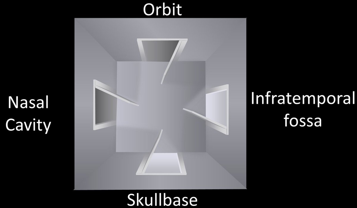
1/Does PTERYGOPALATINE FOSSA anatomy feel as confusing as its spelling? Does it seem to have as many openings as letters in its name?
Let this #tweetorial on PPF #anatomy help you out
#meded #medtwitter #FOAMed #FOAMrad #neurosurgery #neurology #neurorad #neurotwitter #radres
Let this #tweetorial on PPF #anatomy help you out
#meded #medtwitter #FOAMed #FOAMrad #neurosurgery #neurology #neurorad #neurotwitter #radres
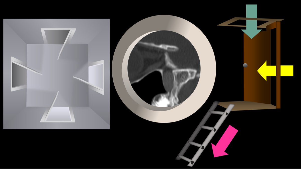
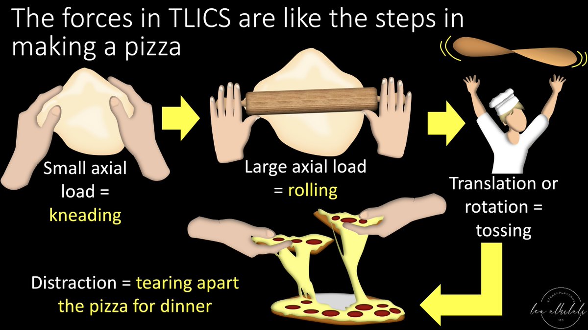
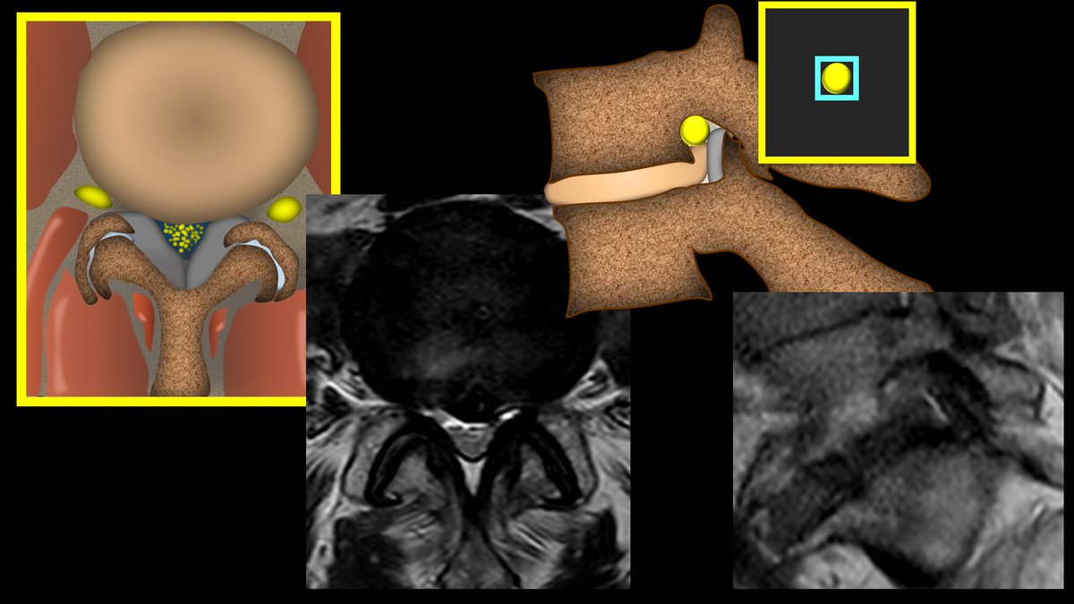
1/Remembering spinal fracture classifications is back breaking work!
A #tweetorial to help your remember the scoring system for thoracic & lumbar fractures—“TLICS” to the cool kids!
#medtwitter #radres #FOAMed #FOAMrad #neurorad #Meded #backpain #spine #Neurosurgery
A #tweetorial to help your remember the scoring system for thoracic & lumbar fractures—“TLICS” to the cool kids!
#medtwitter #radres #FOAMed #FOAMrad #neurorad #Meded #backpain #spine #Neurosurgery

1/Understanding cervical radiculopathy is a pain in the neck! But knowing the distributions can help your search
A #tweetorial to help you remember cervical radicular pain distributions
#medtwitter #radres #FOAMed #FOAMrad #neurorad #Meded #meded #spine #Neurosurgery
A #tweetorial to help you remember cervical radicular pain distributions
#medtwitter #radres #FOAMed #FOAMrad #neurorad #Meded #meded #spine #Neurosurgery

1/ “Say Aaaaaaah!” I was explaining to my resident how I remember tongue anatomy on imaging & he said, “You have to put it on Twitter!”
So here's a #tweetorial about how to remember tongue anatomy on imaging.
#medtwitter #radres #medstudent #FOAMed #FOAMrad #neurorad #meded
So here's a #tweetorial about how to remember tongue anatomy on imaging.
#medtwitter #radres #medstudent #FOAMed #FOAMrad #neurorad #meded
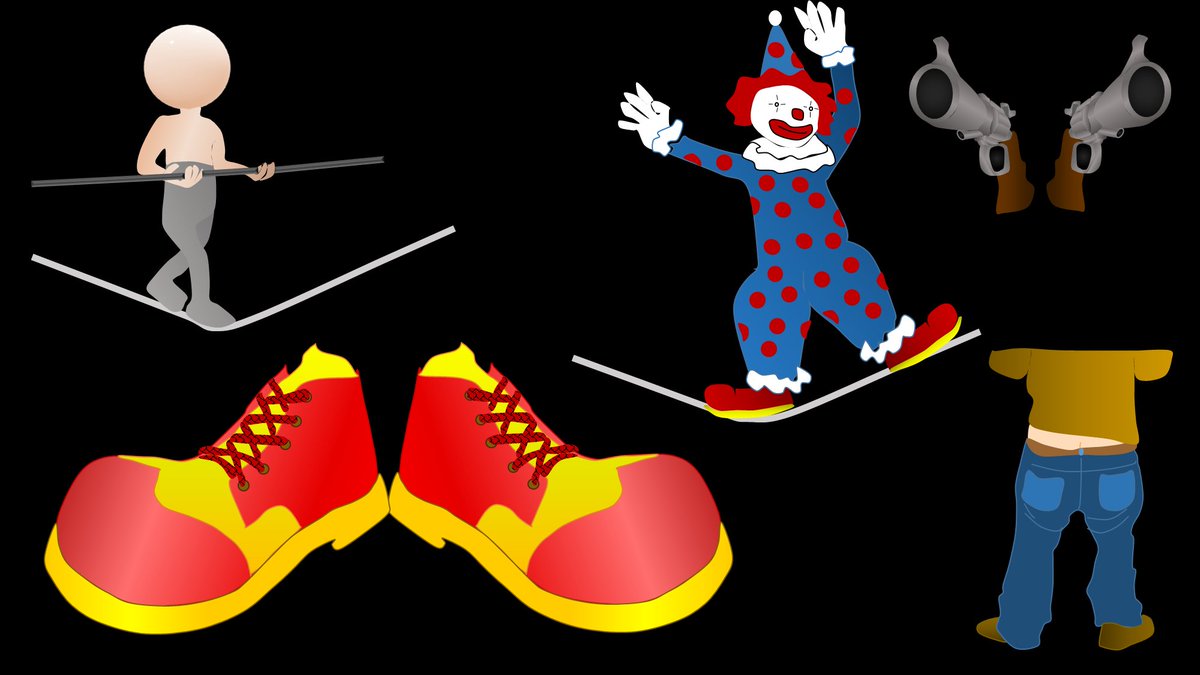
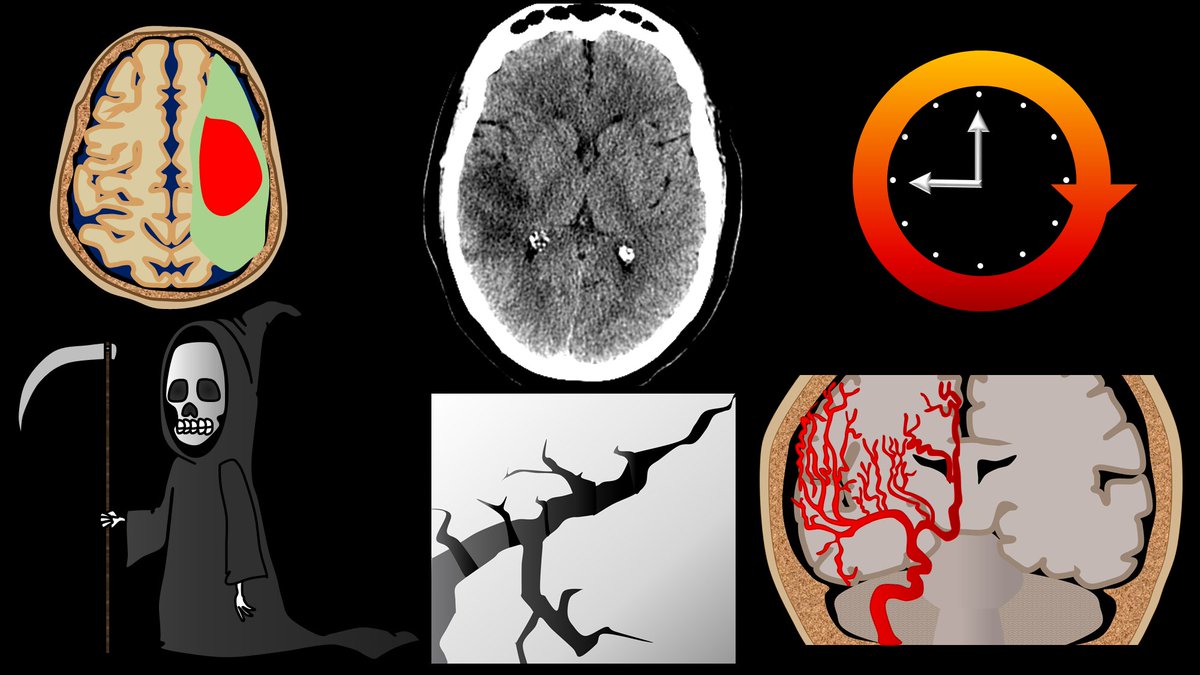
1/Time is brain! So you don’t have time to struggle w/that stroke alert head CT.
Here’s a #tweetorial to help you with the CT findings in acute stroke.
#medtwitter #FOAMed #FOAMrad #ESOC #medstudent #neurorad #radres #meded #radtwitter #stroke #neurology #neurotwitter
Here’s a #tweetorial to help you with the CT findings in acute stroke.
#medtwitter #FOAMed #FOAMrad #ESOC #medstudent #neurorad #radres #meded #radtwitter #stroke #neurology #neurotwitter
1/”Tell me where it hurts.” How back pain radiates can tell you where the lesion is—if you know where to look!
A #tweetorial about how to remember lumbar radicular pain distributions.
#medstudenttwitter #medtwitter #radres #FOAMed #FOAMrad #neurorad #tweetorial #Meded
A #tweetorial about how to remember lumbar radicular pain distributions.
#medstudenttwitter #medtwitter #radres #FOAMed #FOAMrad #neurorad #tweetorial #Meded

1/Do radiologists sound like they are speaking a different language when they talk about MRI? T1 shortening what? T2 prolongation who?
Here’s a translation w/a #tweetorial introduction to MRI.
#medtwitter #FOAMed #FOAMrad #medstudent #neurorad #radres #ASNR23 #neurosurgery
Here’s a translation w/a #tweetorial introduction to MRI.
#medtwitter #FOAMed #FOAMrad #medstudent #neurorad #radres #ASNR23 #neurosurgery

ASNR COTW #143
Hx: Altered mental status with oral pain.
NO SPOILERS!!! Give hints in the form of GIF or answer attached poll.
Answer in 24 hrs
#Neuro #Neurorad #Erad #radres #FOAMed #FOAMrad #medtwitter #ASNRCOTW
Hx: Altered mental status with oral pain.
NO SPOILERS!!! Give hints in the form of GIF or answer attached poll.
Answer in 24 hrs
#Neuro #Neurorad #Erad #radres #FOAMed #FOAMrad #medtwitter #ASNRCOTW
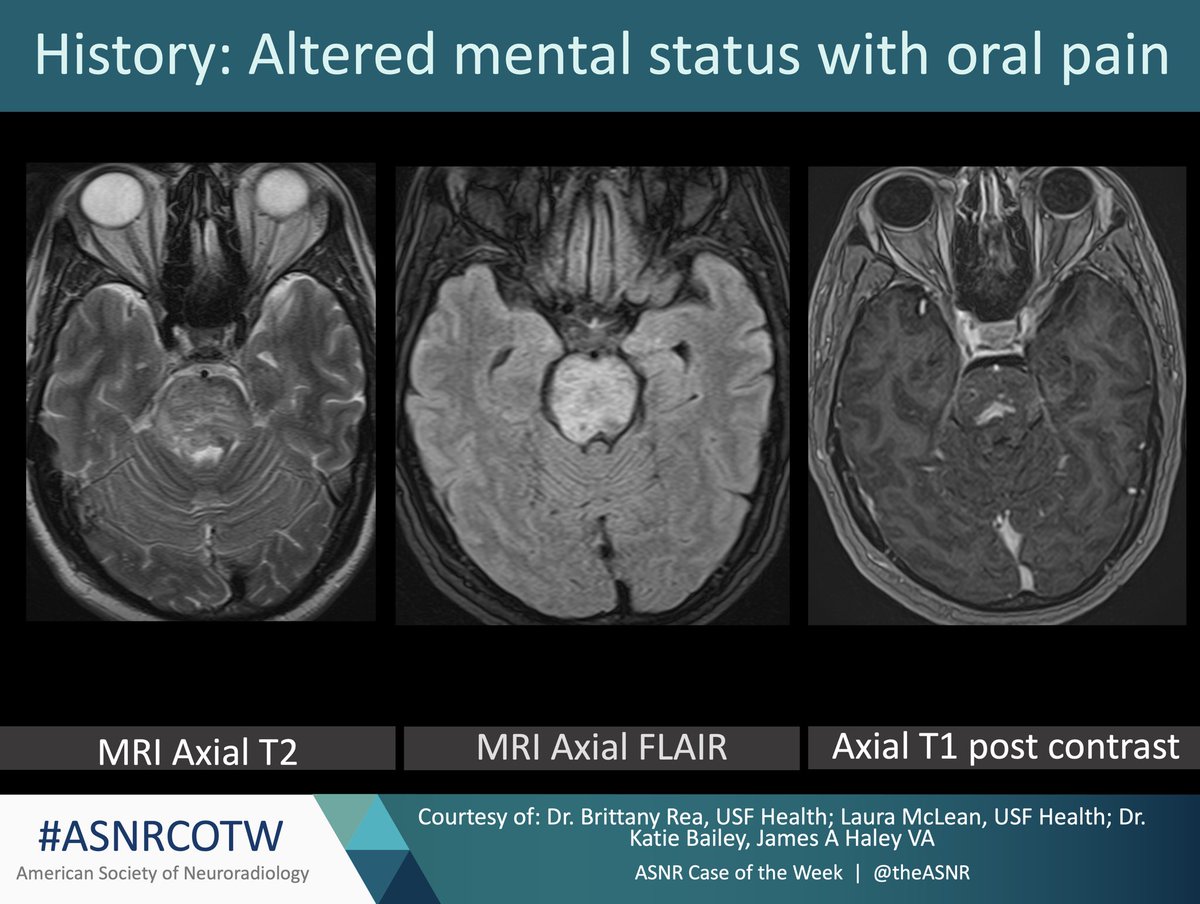
What disease process is associated with oral ulcers and enhancing hyperintense T2 signal lesions in the brain and spinal cord?
ASNR COTW #142
Hx: 62 y/o male initially found unresponsive on the street presents with bradycardia and anterograde amnesia.
NO SPOILERS!!! Give hints in the form of GIF or answer attached poll.
Answer in 24 hrs
#Neuro #Neurorad #radres #FOAMed #FOAMrad #medtwitter #ASNRCOTW
Hx: 62 y/o male initially found unresponsive on the street presents with bradycardia and anterograde amnesia.
NO SPOILERS!!! Give hints in the form of GIF or answer attached poll.
Answer in 24 hrs
#Neuro #Neurorad #radres #FOAMed #FOAMrad #medtwitter #ASNRCOTW

What is the most likely diagnosis given the history and MRI findings in the referenced images?
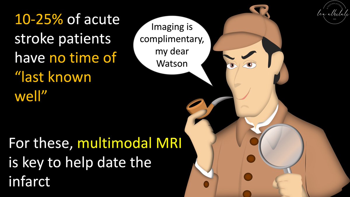
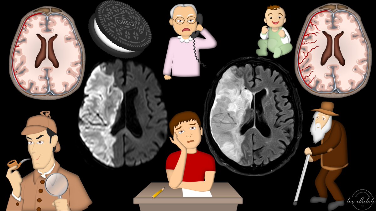
1/Time is brain! But what time is it?
If you don’t know the time of stroke onset, are you able to deduce it from imaging?
Here’s a #tweetorial to help you date a #stroke on MR!
#medtwitter #meded #neurotwitter #neurology #neurorad #radres #radtwitter #radiology #FOAMed #FOAMrad
If you don’t know the time of stroke onset, are you able to deduce it from imaging?
Here’s a #tweetorial to help you date a #stroke on MR!
#medtwitter #meded #neurotwitter #neurology #neurorad #radres #radtwitter #radiology #FOAMed #FOAMrad
80 yo M. Known cardiovascular disease and anemia. Acute abdominal pain and vomit.
Diagnosis? Only ONE answer is correct 😉
#radres #futureradres #FOAMrad #FOAMed #GITwitter #Endoscopy #GIpath
1. Ischemic colitis
2. IBD
3. Tumor
4. None of the above
Diagnosis? Only ONE answer is correct 😉
#radres #futureradres #FOAMrad #FOAMed #GITwitter #Endoscopy #GIpath
1. Ischemic colitis
2. IBD
3. Tumor
4. None of the above
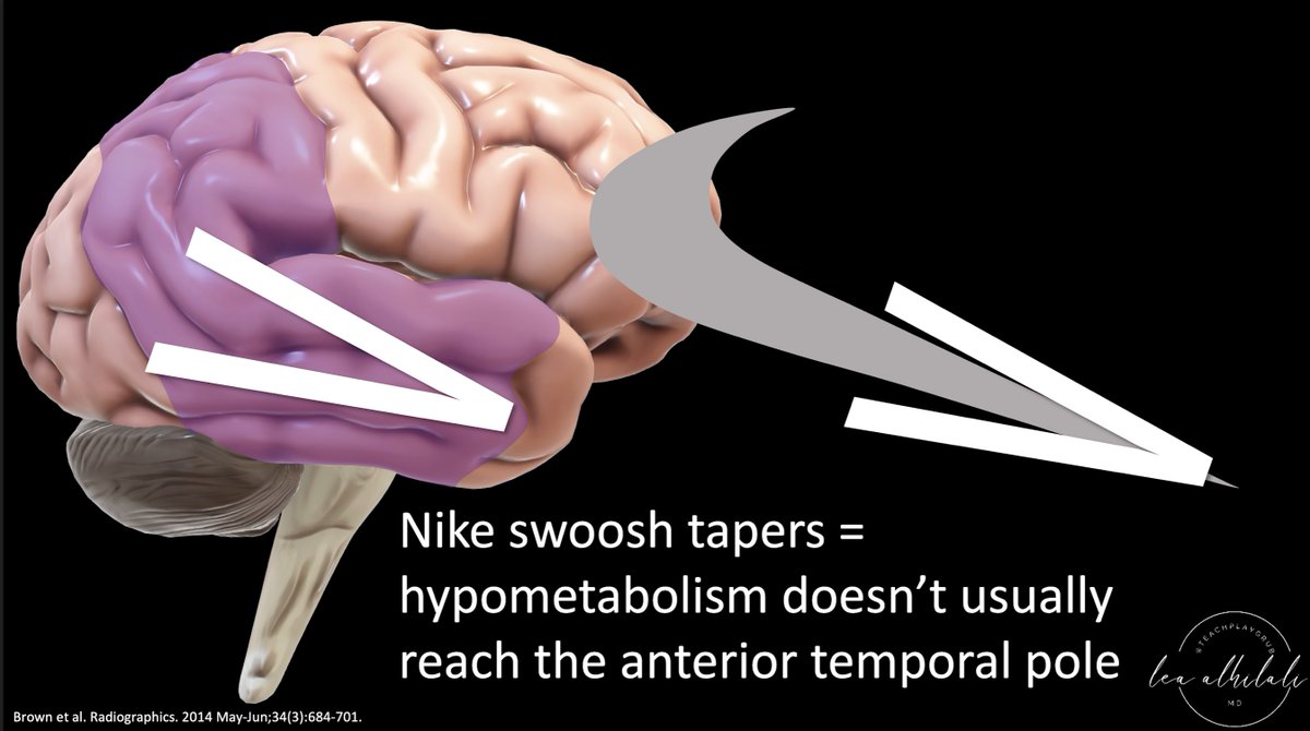
1/Having trouble remembering how to differentiate dementias on imaging?
Here’s a #tweetorial to show you how to remember the imaging findings in dementia & never forget!
#medtwitter #meded #neurorad #radres #dementia #alzheimers #neurotwitter #neurology #FOAMed #FOAMrad #PET
Here’s a #tweetorial to show you how to remember the imaging findings in dementia & never forget!
#medtwitter #meded #neurorad #radres #dementia #alzheimers #neurotwitter #neurology #FOAMed #FOAMrad #PET

1/To be or not 2b?? That is the question!
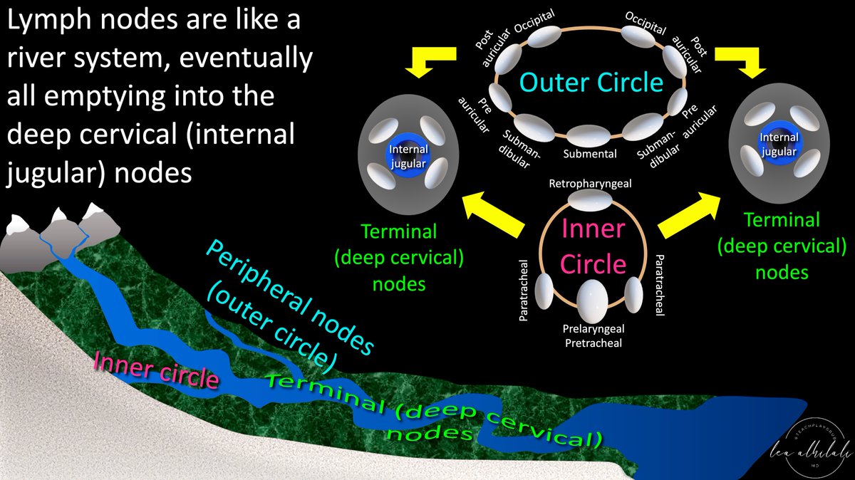
Do you have questions about how to remember cervical lymph node anatomy & levels?
Here’s a #tweetorial to show you how--#Superbowl weekend edition!
#medtwitter #meded #neurorad #HNrad #FOAMed #FOAMrad #radres #radtwitter #ENT #radiology
Do you have questions about how to remember cervical lymph node anatomy & levels?
Here’s a #tweetorial to show you how--#Superbowl weekend edition!
#medtwitter #meded #neurorad #HNrad #FOAMed #FOAMrad #radres #radtwitter #ENT #radiology

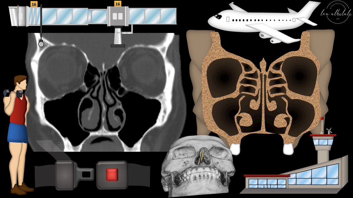
1/Time to FESS up! Do you understand functional endoscopic sinus surgery (FESS)?
If you read sinus CTs, you must know what they’re doing to make the helpful findings
Here’s a #tweetorial to help you!
#medtwitter #meded #FOAMed #FOAMrad #radres #neurorad #HNrad #radtwitter
If you read sinus CTs, you must know what they’re doing to make the helpful findings
Here’s a #tweetorial to help you!
#medtwitter #meded #FOAMed #FOAMrad #radres #neurorad #HNrad #radtwitter
1/”I LOVE spinal cord syndromes!” is a phrase that has NEVER, EVER been said by anyone.
Never fear—here is a #tweetorial on all the incomplete #spinalcord syndromes!
#medtwitter #neurotwitter #neurology #neurosurgery #neurorad #radres #meded #FOAMed #FOAMrad #radtwitter #spine
Never fear—here is a #tweetorial on all the incomplete #spinalcord syndromes!
#medtwitter #neurotwitter #neurology #neurosurgery #neurorad #radres #meded #FOAMed #FOAMrad #radtwitter #spine

1/Have disagreements between radiologists on the degree of cervical canal stenosis become a pain in the neck?!
Here’s a #tweetorial on cervical stenosis grading that’s easy, reproducible & evidence based
#medtwitter #spine #neurosurgery #radres #neurorad #meded #FOAMed #FOAMrad
Here’s a #tweetorial on cervical stenosis grading that’s easy, reproducible & evidence based
#medtwitter #spine #neurosurgery #radres #neurorad #meded #FOAMed #FOAMrad

1/Tonsillar or peritonsillar abscess? That is the question! When you look at a neck CT, do you know which one to say?
A #tweetorial on #tonsillitis complications
#medtwitter #radtwitter #neurorad #radres #meded #FOAMed #FOAMrad #HNrad #medstudenttwitter #RSNA2022
A #tweetorial on #tonsillitis complications
#medtwitter #radtwitter #neurorad #radres #meded #FOAMed #FOAMrad #HNrad #medstudenttwitter #RSNA2022

1/They say form follows function! Brain #MRI anatomy is best understood in terms of both form & function
A #tweetorial on how to remember important functional #brain #anatomy
#meded #medtwitter #neurosurgery #neurology #neurorad #FOAMed #FOAMrad #radiology #medstudent #radres
A #tweetorial on how to remember important functional #brain #anatomy
#meded #medtwitter #neurosurgery #neurology #neurorad #FOAMed #FOAMrad #radiology #medstudent #radres

🌓🤪 Pac-Man ate my visual fields!
50 year old person presents with sudden onset headache and seeing "pieces of things" on the R.
Visual fields look like this👇👇
➡️What's happening?
#MedTwitter #MedStudentTwitter #neurotwitter #MedEd #Neurology #EndNeurophobia #stroke
50 year old person presents with sudden onset headache and seeing "pieces of things" on the R.
Visual fields look like this👇👇
➡️What's happening?
#MedTwitter #MedStudentTwitter #neurotwitter #MedEd #Neurology #EndNeurophobia #stroke

🗺️🧠 Where across the visual system is likely the lesion?
#Ophthalmology
#Ophthalmology
⭕️ Which artery has to be implicated, if this presentation is due to an ischemic #stroke and the rest of the neuro exam is normal?
1/Hate it when one radiologist called the stenosis mild, the next one said moderate--but it was unchanged?!
Here’s a #tweetorial of a lumbar grading system that’s easy, reproducible & evidence-based
#medtwitter #spine #neurosurgery #radres #neurorad #meded #FOAMed #FOAMrad
Here’s a #tweetorial of a lumbar grading system that’s easy, reproducible & evidence-based
#medtwitter #spine #neurosurgery #radres #neurorad #meded #FOAMed #FOAMrad
Multiple Sclerosis - what to look for on MRI! A quick overview. First slide shows Jean-Martin Charcot, one of the fathers of Neurology (the painting is a bit outdated to say the least, but interesting from a historical point of view). #radres #neurorad #FOAMrad #FOAMed 
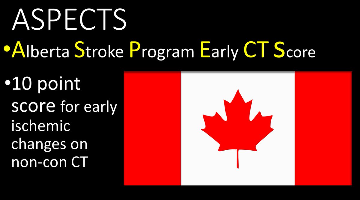
1/One important aspect to stroke care is well... ASPECTS.😂
Here’s a #tweetorial to help you remember this basic #STROKE scoring system
#medtwitter #FOAMed #FOAMrad #medstudenttwitter #medstudent #neurorad #radiology #radres #neurology #Neurosurgery #CT #meded #neurotwitter
Here’s a #tweetorial to help you remember this basic #STROKE scoring system
#medtwitter #FOAMed #FOAMrad #medstudenttwitter #medstudent #neurorad #radiology #radres #neurology #Neurosurgery #CT #meded #neurotwitter

Cystic supratentorial brain tumors in adults, the usual and the unusual. Non-exhaustive list of differential diagnostic considerations. The PA had a nice mural nodule, not shown on this figure. #radres #neurorad #FOAMrad #FOAMed #MedEd #RadEd 

impossible to tell which is which on only T1-images with GD. Whenever I see an intra-axial tumor in an adult, I automatically consider GBM and metastases as they are the most frequent brain tumors in adults, so even when the tumor looks strange or atypical, they are a good bet.
Brain abcess and epidermoid cyst are pretty straightforward on diffusion-weighted images as their content is very diffusion restrictive. Brain abscess has a thick enhancing capsule, epidermoidcyst doesn't.


