Discover and read the best of Twitter Threads about #ecg
Most recents (24)
1) Welcome to Part 1 of a new #accredited #tweetorial in our series of educational programs on #hypertrophic #cardiomyopathy #HCM. Previous programs, still available for 🆓CE/#CME, are at cardiometabolic-ce.com/category/hcm/.
Now you can earn another 1.5hr credit by following ALL of this 🧵!
Now you can earn another 1.5hr credit by following ALL of this 🧵!
2) Our expert author is Sergio Kaiser MD PhD FACC FESC 🇧🇷🇮🇱 @pabeda1, cardiologist 🫀, Professor 🎓 of #InternalMedicine, Rio de Janeiro State University. He brings the general cardiologist's perspective to our #HCM discussions. Read and learn!
#FOAMed #CardioTwitter
#FOAMed #CardioTwitter

3) This program is supported by an unrestricted educational grant from Bristol Myers Squibb. Statement of accreditation and faculty disclosures at cardiometabolic-ce.com/disclosures/. Credit for #physicians #nursepractitioners #physicianassociates #nurses #pharmacists from @academiccme.
When your ER colleague calls you to have a quick look on an #ecg … the 28 yo pat reports frequent episodes of palpitations since childhood that he can usually terminate with deep breathing. #Epeeps @EPeeps_Bot #CardioTwitter know what’s going on. Do’s and Don’t’s? twitter.com/i/web/status/1…
10 min after giving 2mg/kg body weight Flecainid iv. twitter.com/i/web/status/1…
1/ Let’s talk about focal atrial tachycardia #CardioTwitter.
🧵👇 by @ekgdx
#ekgdx #ecg #ekg #MedTwitter #medicine #medstudents #basic #education #graphic
🧵👇 by @ekgdx
#ekgdx #ecg #ekg #MedTwitter #medicine #medstudents #basic #education #graphic

2/ Focal atrial tachycardia is characterized by at least three or more consecutive ectopic P waves with similar morphology, usually arising from a single ectopic focus.
#ekgdx
#ekgdx
3/ Criteria
✅ ≥3 consecutive similar ectopic P waves (usually inverted in inferior leads).
✅ Atrial rate >100 bpm.
✅ QRS usually narrow unless pre-existing BBB or aberrant conduction.
#ekgdx
✅ ≥3 consecutive similar ectopic P waves (usually inverted in inferior leads).
✅ Atrial rate >100 bpm.
✅ QRS usually narrow unless pre-existing BBB or aberrant conduction.
#ekgdx
A young man was found down in cardiac arrest. 911 was called and paramedics achieved ROSC en route prior to arrival in the ER. No further history was available. This #ECG was recorded.
What’s the diagnosis?
What’s the diagnosis?

If you’re a new follower I always post the answer with explanation the next day. If you’re new to my account—follow me if you want to learn about medical emergencies
Answer:
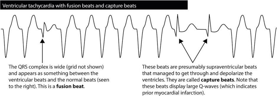
AV dissociation, capture beats and fusion beats are all hallmarks of ventricular tachycardia
A simple understanding 👇
#CardioTwitter #Cardiology #Medtwitter
A simple understanding 👇
#CardioTwitter #Cardiology #Medtwitter
What’s a “capture beat” ?
During ventricular tachycardia, sometimes the SA nodal firing takes control of independent ventricular depolarization for brief moment, as this happens, “CAPTURE BEATS” appear. They are nothing but a brief moment of normal looking P-QRS pattern
During ventricular tachycardia, sometimes the SA nodal firing takes control of independent ventricular depolarization for brief moment, as this happens, “CAPTURE BEATS” appear. They are nothing but a brief moment of normal looking P-QRS pattern

Whats a “fusion beat” ?
During ventricular tachycardia, sometimes the SA nodal impulse and the independent ventricular impulse combine together to create a mix looking P-QRS pattern called fusion beats.
#ECG @EPeeps_Bot #ecglearning #CardiacArrest

During ventricular tachycardia, sometimes the SA nodal impulse and the independent ventricular impulse combine together to create a mix looking P-QRS pattern called fusion beats.
#ECG @EPeeps_Bot #ecglearning #CardiacArrest


1/ Let’s talk about T waves #CardioTwitter
The T wave represents typically ventricular repolarization. It is the most labile wave on the EKG surface.
Normal T wave
✅ Morphology: Asymmetric.
✅ Amplitude: ≤6 mm in limb leads and ≤10 mm in precordial leads.
#MedTwitter
The T wave represents typically ventricular repolarization. It is the most labile wave on the EKG surface.
Normal T wave
✅ Morphology: Asymmetric.
✅ Amplitude: ≤6 mm in limb leads and ≤10 mm in precordial leads.
#MedTwitter

2/ Tall upright T wave
Tall upright T waves are usually characterized by tall and peaked shape.
✅ Amplitude: >6 mm in limb leads and >10 mm in precordial leads.
Causes: Hyperkalemia, hyperacute MI, normal variant, prinzmetal angina, aortic stenosis, LVH, RVH, others.
#ecg
Tall upright T waves are usually characterized by tall and peaked shape.
✅ Amplitude: >6 mm in limb leads and >10 mm in precordial leads.
Causes: Hyperkalemia, hyperacute MI, normal variant, prinzmetal angina, aortic stenosis, LVH, RVH, others.
#ecg

3/ Notched T wave
Possible causes: May be caused by morphological changes in the cardiomyocytes' action potential waveforms. Another causes include: Drugs (such as Dofetilide, Quinidine, Ranolazine, Verapamil), long QT syndrome, athletes, others.
#MedTwitter #MedStudentTwitter
Possible causes: May be caused by morphological changes in the cardiomyocytes' action potential waveforms. Another causes include: Drugs (such as Dofetilide, Quinidine, Ranolazine, Verapamil), long QT syndrome, athletes, others.
#MedTwitter #MedStudentTwitter

Are you a #juniordoctor or #medstudent?
Here's 10 great FREE modules to help get you started on the wards!
#meded #medschool #tipsfornewdocs #juniordocs #FOAMed #medtwitter #medstudenttwitter #juniordoctors #medstudents
Here's 10 great FREE modules to help get you started on the wards!
#meded #medschool #tipsfornewdocs #juniordocs #FOAMed #medtwitter #medstudenttwitter #juniordoctors #medstudents
Occasionally you'll need to perform sterile procedures. Make sure you prepare the best you can
osler.app.link/CztXIRjyntb
osler.app.link/CztXIRjyntb

Providing basic life support is a core skill for all healthcare staff
osler.app.link/u4fe8uqyntb
#basiclifesupport #bls #FOAMresus #resuscitation
osler.app.link/u4fe8uqyntb
#basiclifesupport #bls #FOAMresus #resuscitation

1/ Let’s talk about the ST segment #CardioTwitter.
ST segment normally represents the interval between ventricular depolarization and repolarization.
Normal ST
✅ Usually isolectric or may vary from 0.5 mm below to 1 mm above isolectric line in L leads.
#ekgdx #Medicine
ST segment normally represents the interval between ventricular depolarization and repolarization.
Normal ST
✅ Usually isolectric or may vary from 0.5 mm below to 1 mm above isolectric line in L leads.
#ekgdx #Medicine

2/ ST elevation (STE)
ST changes suggesting myocardial injury:
✅ New STE ≥1 mm in all leads other than V2 or V3.
✅ New STE in V2-V3 ≥2 mm in men older than 40 years old and ≥2.5 mm in men younger than 40 years old or ≥1.5 mm in women.
#ekgdx #Medstudent #MedTwitter
ST changes suggesting myocardial injury:
✅ New STE ≥1 mm in all leads other than V2 or V3.
✅ New STE in V2-V3 ≥2 mm in men older than 40 years old and ≥2.5 mm in men younger than 40 years old or ≥1.5 mm in women.
#ekgdx #Medstudent #MedTwitter

3/ Types of ST segment elevation include:
✅ Convex Upward (previous pic)
✅ Horizontal (this one)
✅ Concave Upward (see next)
✅ Obliquely Straight (see next)
#ekgdx #st #MedTwitter #ecg #ekg #CardioTwitter #Medstudent
✅ Convex Upward (previous pic)
✅ Horizontal (this one)
✅ Concave Upward (see next)
✅ Obliquely Straight (see next)
#ekgdx #st #MedTwitter #ecg #ekg #CardioTwitter #Medstudent

Three #cardiology cases with diagnostic ECGs in our resus room today and some learning points for emergency clinicians
#ecg #ekg
#ecg #ekg
We suspected this was atrial flutter
Rather than subject a patient to the horror of iv adenosine (which only reveals flutter - it can’t convert it), we moved the ECG limb leads around to get a ‘Lewis lead’ which better shows atrial activity
(See litfl.com/lewis-lead-s5-… )
Rather than subject a patient to the horror of iv adenosine (which only reveals flutter - it can’t convert it), we moved the ECG limb leads around to get a ‘Lewis lead’ which better shows atrial activity
(See litfl.com/lewis-lead-s5-… )

1/ Let’s talk about PR Interval - Segment #CardioTwitter.
The PR interval represents the time between the onset of atrial depolarization and the onset of ventricular depolarization and reflects conduction through the AV node.
🧵by @ekgdx
The PR interval represents the time between the onset of atrial depolarization and the onset of ventricular depolarization and reflects conduction through the AV node.
🧵by @ekgdx

2/ PR Interval
Criteria
✅ Normal PR interval: 0.12 - 0.20 sec.
✅ Prolonged PR interval: >0.20 sec.
✅ Short PR interval: <0.12 sec.
The term “PQ interval” is preferred by some EKG lover because it is the period actually measured unless the Q wave is absent.
#PR #interval
Criteria
✅ Normal PR interval: 0.12 - 0.20 sec.
✅ Prolonged PR interval: >0.20 sec.
✅ Short PR interval: <0.12 sec.
The term “PQ interval” is preferred by some EKG lover because it is the period actually measured unless the Q wave is absent.
#PR #interval
3/ The PR segment is the segment between the end of the P wave and the start of the QRS complex.
Criteria
✅ Normal PR segment: Usually isolectric.
✅ PR segment elevation: ≥0.5 mm.
PR segment elevation causes: Atrial ischaemia/infarction, myopericarditis, PE, others.
#ekgdx
Criteria
✅ Normal PR segment: Usually isolectric.
✅ PR segment elevation: ≥0.5 mm.
PR segment elevation causes: Atrial ischaemia/infarction, myopericarditis, PE, others.
#ekgdx

🆘59 year male smoker
k/c/o COPD, DM, HTN with DCMP (EF=20%) palpitations and dizziness x 2 days
O/E : B/L crepts +, rest
ECG👇
#Medtwitter #MedEd2022 #ECG #CardioEd
k/c/o COPD, DM, HTN with DCMP (EF=20%) palpitations and dizziness x 2 days
O/E : B/L crepts +, rest
ECG👇
#Medtwitter #MedEd2022 #ECG #CardioEd

What is the likely rhythm ?
1/ Let’s talk about P waves #CardioTwitter.
The P wave is the first positive deflection on the EKG and represents atrial depolarization. The first half represents right atrial depolarization and the second half represents left atrial depolarization.
By @ekgdx
The P wave is the first positive deflection on the EKG and represents atrial depolarization. The first half represents right atrial depolarization and the second half represents left atrial depolarization.
By @ekgdx

2/ Normal P wave
Criteria
✅ Axis: 0° to +75°.
✅ Amplitude (L leads): <2.5 mm.
✅ Amplitude (P leads): <1.5 mm.
✅ Duration: 0.08 - 0.11 sec.
✅ Morphology: Upright in I, II, aVF and inverted in aVR.
#Pwaves #ecg #ekg #ekgdx #medicine #MedTwitter #MedStudentTwitter #basic
Criteria
✅ Axis: 0° to +75°.
✅ Amplitude (L leads): <2.5 mm.
✅ Amplitude (P leads): <1.5 mm.
✅ Duration: 0.08 - 0.11 sec.
✅ Morphology: Upright in I, II, aVF and inverted in aVR.
#Pwaves #ecg #ekg #ekgdx #medicine #MedTwitter #MedStudentTwitter #basic
Acute pulmonary embolism (PE) is one of the most serious form of venous thromboembolism. The clinical presentation of PE is variable and often nonspecific making the diagnosis challenging.
1/
#CardioTwitter
1/
#CardioTwitter

Criteria
✅ Sinus tachycardia (most common).
✅ S1Q3T3 pattern (may be present up to 30% of cases).
✅ Simultaneous T wave inversions in the inferior leads and right precordial leads can be seen.
✅ Right axis deviation.
✅ RBBB (complete or incomplete).
2/
#MedTwitter #ecg
✅ Sinus tachycardia (most common).
✅ S1Q3T3 pattern (may be present up to 30% of cases).
✅ Simultaneous T wave inversions in the inferior leads and right precordial leads can be seen.
✅ Right axis deviation.
✅ RBBB (complete or incomplete).
2/
#MedTwitter #ecg
Here’s a very important #ECG of a 60-year-old lady who presented with chest pain and shortness of breath that began while walking her dog. She was also COVID+
What’s the diagnosis?
What’s the diagnosis?

If you’re a new follower I always post the answer with explanation the next day. If you’re new to my account—follow me if you want to learn about medical emergencies
Here’s a very important #ECG that was recorded in a 50-year-old lady shortly before she suddenly went into cardiac arrest
What’s the diagnosis?
What’s the diagnosis?

If you’re a new follower I always post the answer with explanation the next day. If you’re new to my account—follow me if you want to learn about medical emergencies
Its WPW pattern --> Derpak Sir has provided a methology to localize the Kent bundle --> I can't call it WPW syndrome for there is no hx of pre excited NCT.
*Deepak Sir
My fiancé and I were supposed to get married in July 2022. Given the first 2 vaccine mandates, we figured that we had no choice but to keep on taking the boosters in order to be able to travel for our honeymoon / have liberties to go to stores etc.
We decided to take the 3rd @moderna_tx booster anticipating that perhaps it would also be mandated by the #Canadian #government.
Here’s the #ECG of a 68 year old man who was rushed to the ER by paramedics
BP: 80/40
HR: 150
RR: 35
SPO2: 95%
What’s the diagnosis?
BP: 80/40
HR: 150
RR: 35
SPO2: 95%
What’s the diagnosis?

Answer: HYPERKALEMIA
There’s a wide complex tachycardia with RBBB morphology. There are features here concerning for several life-threatening diagnoses including: V-Tach, Pulmonary Embolism, Acute Coronary Occlusion. But ALL these changes resolved with empiric ↑K+ treatment…
There’s a wide complex tachycardia with RBBB morphology. There are features here concerning for several life-threatening diagnoses including: V-Tach, Pulmonary Embolism, Acute Coronary Occlusion. But ALL these changes resolved with empiric ↑K+ treatment…
🧵 1/7 Ever wondered why the Osborn wave looks the way it does? Stay with me during my newest #tweetorial. A thread 🧵1/7
#cardiotwitter #EPeeps #CardioEd #MedTwitter @TRassafMD @YoungDgk @DGK_org @YoungDZHK @AaronGoodman33 @Steph_Achenbach @fuzzymittens @AvrahamCooperMD
#cardiotwitter #EPeeps #CardioEd #MedTwitter @TRassafMD @YoungDgk @DGK_org @YoungDZHK @AaronGoodman33 @Steph_Achenbach @fuzzymittens @AvrahamCooperMD

2/7 History
First described in 1953 by Osborn (camel-hump sign) upon #hypothermia in dogs. Upon systemic analysis similar #ECG patterns have been described in
➡️ hypercalcemia
➡️ brain injury
➡️ SAB
➡️ vasospastic angina / ischemia
First described in 1953 by Osborn (camel-hump sign) upon #hypothermia in dogs. Upon systemic analysis similar #ECG patterns have been described in
➡️ hypercalcemia
➡️ brain injury
➡️ SAB
➡️ vasospastic angina / ischemia
3/7 Emslie-Smith et al showed that Osborn waves manifested more in epicardial than endocardial leads. Others finally showed that 4-aminopyridine sensitive transient outward current (Ito) is responsible and predominantly located in epicardium. ⬇️ heart rate led to ⬆️ Ito current
Here’s an incredible #ECG recorded in a 60 year old man with chest pain and palpitations
What’s the diagnosis?
What’s the diagnosis?

If you’re a new follower I always post the answer with explanation the next day. If you’re new to my account—follow me if you want to learn about Emergency ECGs
My 1st symptoms of #COVID were 7 weeks ago tomorrow.
Yesterday I went for an #ECG b/c I'm having heart symptoms that weren't there before #COVID19.
I'm also having issues with my blood sugar which weren't there before #Covid_19
The technician said he's doing several ECG's ++
Yesterday I went for an #ECG b/c I'm having heart symptoms that weren't there before #COVID19.
I'm also having issues with my blood sugar which weren't there before #Covid_19
The technician said he's doing several ECG's ++
daily on people who are having heart complications due to #COVID19.
I'm on leave from my job. And now I'm in need of health care I wasn't before. I'm scared of what is happening inside my body and brain.
@TimHoustonNS got rid of all the means that protected the #NovaScotia ++
I'm on leave from my job. And now I'm in need of health care I wasn't before. I'm scared of what is happening inside my body and brain.
@TimHoustonNS got rid of all the means that protected the #NovaScotia ++
population. Our health care and workers are crumbling under the weight of it all.
Businesses are closing. Housing is a mess. The climate is in shambles.
What are we going to do?
#nspoli #covid19NS #CovidIsNotOver #COVID19 #COVID
Businesses are closing. Housing is a mess. The climate is in shambles.
What are we going to do?
#nspoli #covid19NS #CovidIsNotOver #COVID19 #COVID
Here’s an important #ECG that was recorded in a 50-year-old man just 15 minutes before he suddenly went into pulseless V-Tach
What’s the diagnosis?
What’s the diagnosis?

This was an unbelievable case. If you’re a new follower I always post the answer with explanation the next day. If you’re new to my account—follow me if you want to learn about Emergency ECGs
Hey #OhGottPJ - Ihr seid alleine in der ZNA und seht diesen Monitorausschnitt - was passiert da? Und was jetzt? :D
#ekg #ecg #cardiotwitter
#ekg #ecg #cardiotwitter

Man sieht irgendwas, was zu einer Breitkomplextachykardie umspringt in der Monitorüberwachung.
1. Wir sind uns ja relativ einig es handelt sich um einen potentiellen Notfall also:
In case of emergency, take your own pulse first!
1. Wir sind uns ja relativ einig es handelt sich um einen potentiellen Notfall also:
In case of emergency, take your own pulse first!
2. Hilfe rufen (alleine ist blöd)
Here’s a great #ECG of a 60 year old man with chest pain and shortness of breath. This is a fantastic case—with a twist!
What’s the diagnosis?
What’s the diagnosis?

If you’re a new follower I always post the answer with explanation the next day. If you’re new to my account, follow me if you want to learn about Emergency ECGs
Here’s a video I made breaking down this rare and fascinating case of a 60 year old man with chest pain and shortness of breath
#FOAMed
#FOAMed


