Discover and read the best of Twitter Threads about #cxr
Most recents (5)
What is the cause of acute and #severe_respiratory_distress?
@BrownJHM @grepmeded #MedTwitter #cxr #respiratory #distress
@BrownJHM @grepmeded #MedTwitter #cxr #respiratory #distress

What is the cause of acute and #severe_respiratory_distress?
No history of #trauma
@BrownJHM
@grepmeded
#MedTwitter #cxr #respiratory #distress
No history of #trauma
@BrownJHM
@grepmeded
#MedTwitter #cxr #respiratory #distress
What is the most important step in mangement ?
1) Welcome to a new #accredited #tweetorial on #Bronchiectasis (#NCFB or #bronchiectasis) & its management, by Christina Thornton MD (@Cthornton32), respirologist & clinician scientist in Calgary 🇨🇦. Follow along and earn 0.75h CE/#CME #physicians #nurses #NPs #PAs #pharmacists! 

2) I am very excited to be among the founding faculty in this initiative! FOLLOW US for awesome expert-led education #pulmtwitter!
👍@BronchiectasisR @COPDFoundation @EMBARCnetwork @ELF @profJDchalmers @sunjayMD @DrHollyKeir @becleartoday @ephesians_1_7 @NTMinfo @AlibertiStefano
👍@BronchiectasisR @COPDFoundation @EMBARCnetwork @ELF @profJDchalmers @sunjayMD @DrHollyKeir @becleartoday @ephesians_1_7 @NTMinfo @AlibertiStefano
3) This program is supported by an educational grant from Insmed & is intended for healthcare professionals. Statement of accreditation and faculty disclosures at pulmonarymed-ce.com/disclosures/. CE/#CME credit from @academiccme.
Lets understand the basic principles of chest x-ray interpretation (1/10)
Use the inside-out approach. Start from inside most structures and then go outward (2/10)
Look at trachea, mediastinum, cardiac size & borders. Don't forget looking at the hilum (3/10)
Non-COVID teaching: Can you identify this unusual cause of chronic cough? (Hint: always carefully look at tubes and lines on #CXR and axis on #EKG) #medtwitter #FOAMed #FOAMrad 


So let’s jump into this #FOAMed case. On the CXR we’ve got a couple interesting things:
* the PICC line appears to terminate on the wrong (e.g. left) side (white arrow)
* the aortic knob is on the wrong (e.g. right) side (grey arrow)
* it’s hard to identify the cardiac silhouette

* the PICC line appears to terminate on the wrong (e.g. left) side (white arrow)
* the aortic knob is on the wrong (e.g. right) side (grey arrow)
* it’s hard to identify the cardiac silhouette


Some of these findings are easy to miss. To avoid missing things, it’s really important to have a systematic approach to reading a CXR. Here’s a good one:
➡️mededportal.org/doi/10.15766/m…
➡️mededportal.org/doi/10.15766/m…
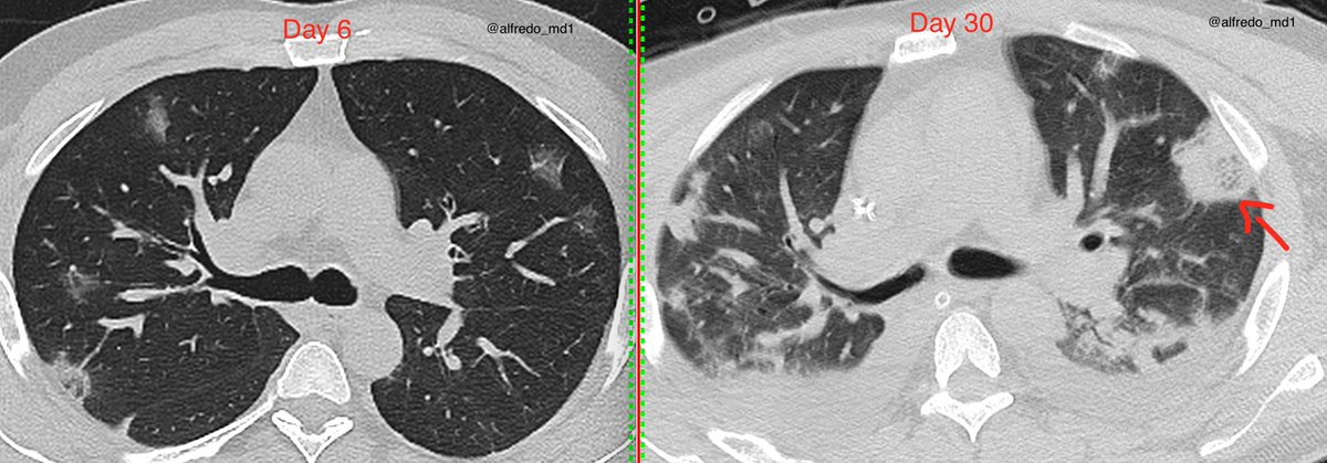
39yo male, BMI=31, no other risk factors. Fever and myalgia started 6 days ago. #COVID19 (+). Tx with steroids & tocilizumab during hospital stay. Day 19➡️Galactomannan antigen positive (Aspergillus). #CT on day 6 with classic #COVID19 pattern. 1/4 


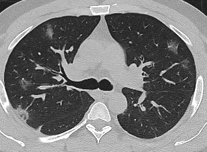


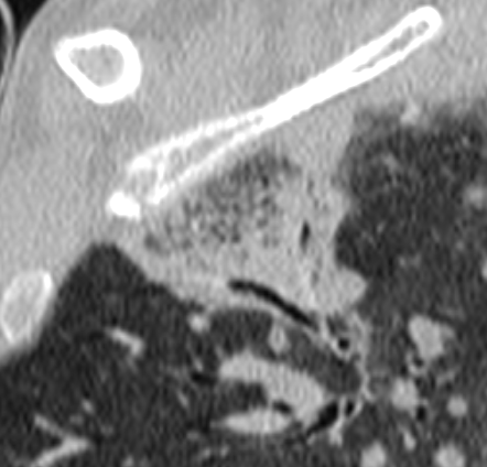
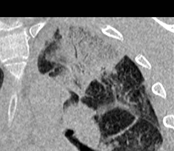
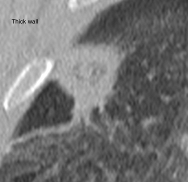
...#CT on day 30 shows 3 rounded lesions with a thick wall in the upper lobes not present on initial CT, suggestive of invasive Aspergillosis. Only risk factors are immunosuppressant drugs used during hospital stay. Next step ➡️ Biopsy?? ... 2/4 


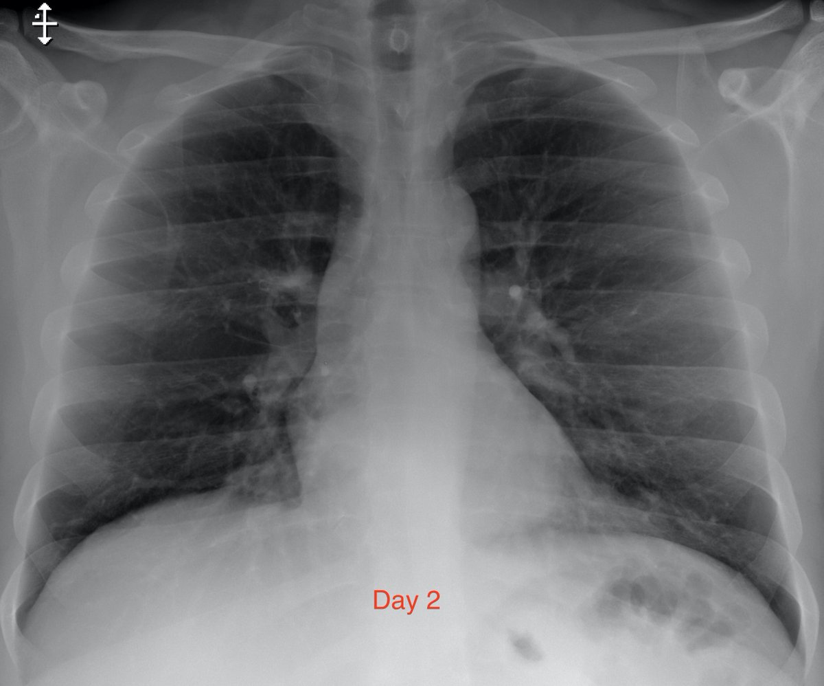
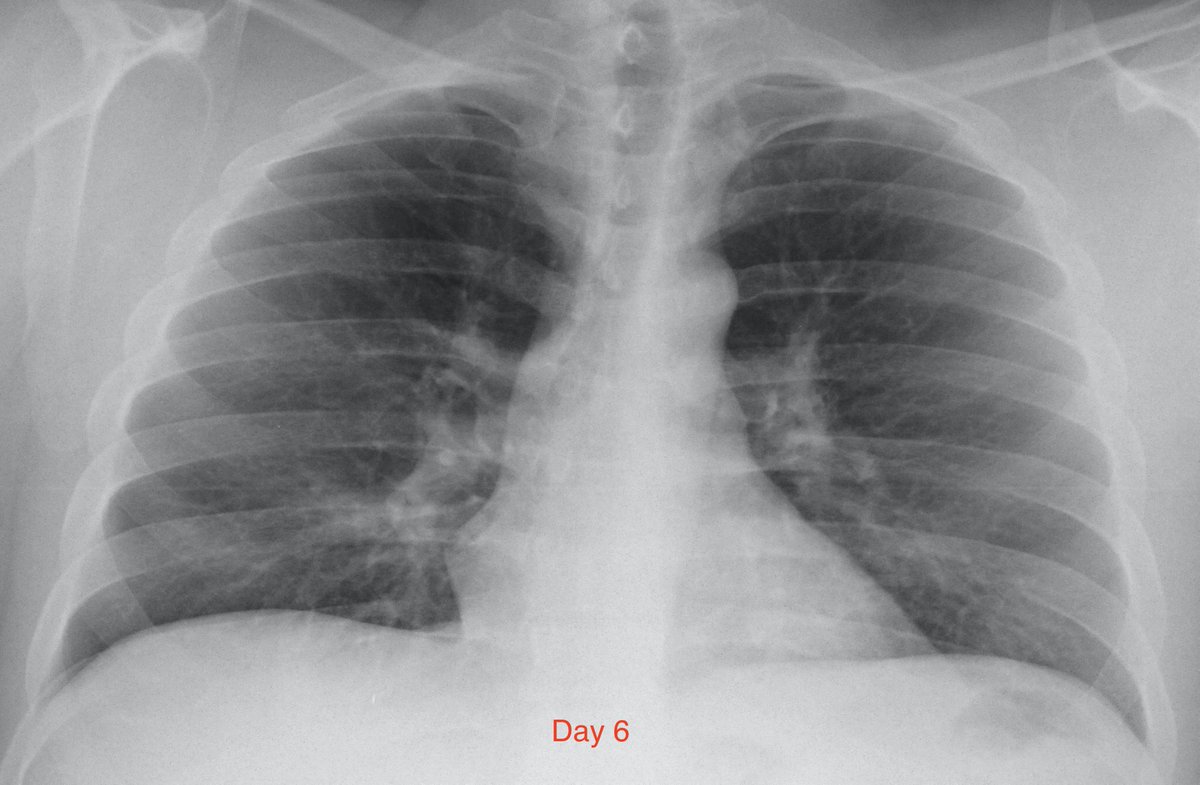
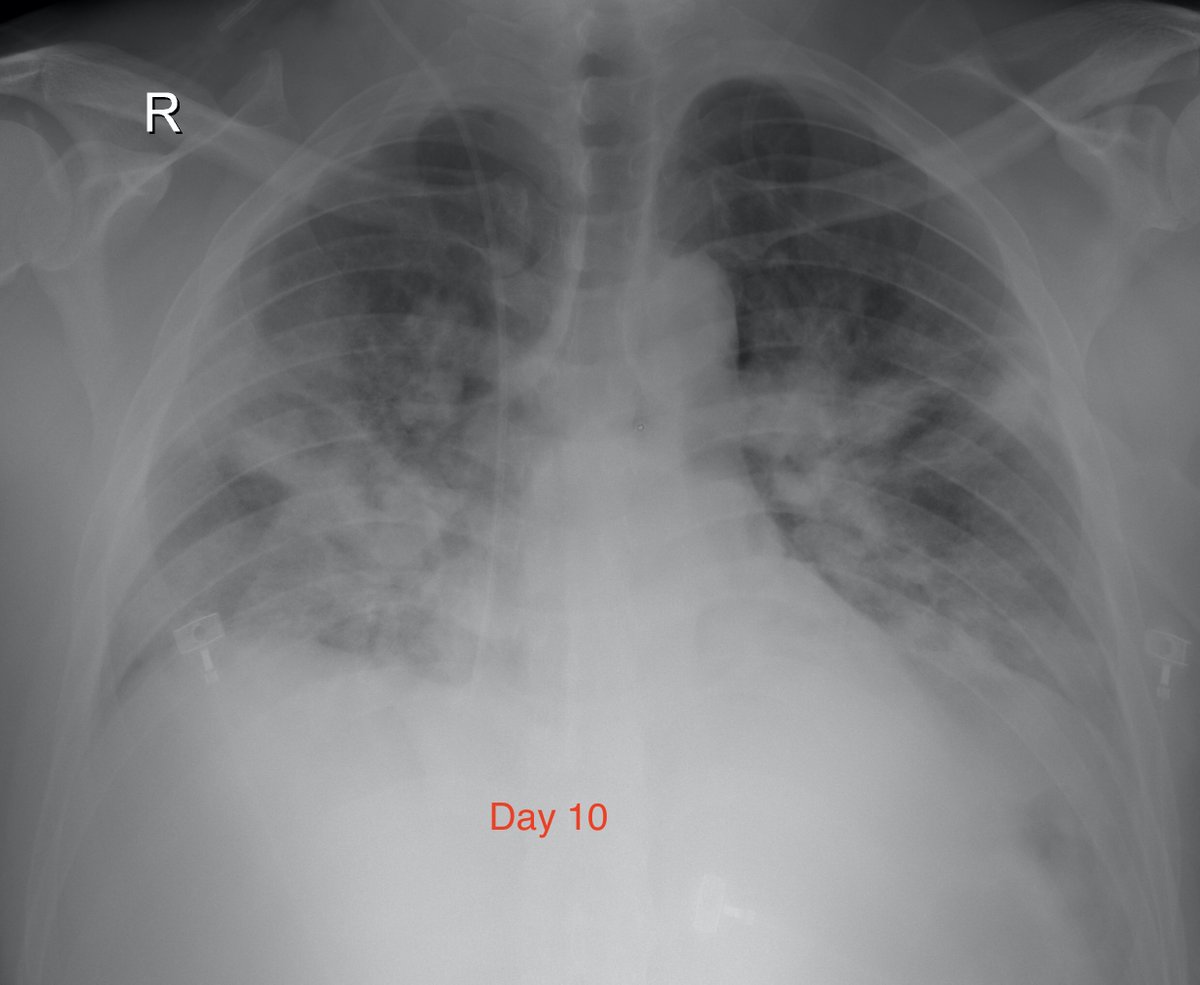
... Here are the #CXR at day 2,6,10 and 14 after the onset of symptoms. Notice how CXR is normal on day 2 and significantly worse on day 10 (peak stage of symptoms). 3/4 


