Discover and read the best of Twitter Threads about #VExUS
Most recents (24)
#POCUS enthusiasts, is this IVC normal, abnormal? (asking about possibilities, not definitive conclusions)
#FOAMcc #MedEd #Nephpearls
(Transverse view in thread)
#FOAMcc #MedEd #Nephpearls
(Transverse view in thread)
IVC transverse #POCUS
#POCUS answer:
I deliberately avoided clinical context in the original tweet to gather multiple opinions.
Image obtained from a thin, trained athlete with resting HR in 40s-50s.
A dilated IVC is commonly seen in this setting with no right heart pathology. In addition, particulate… twitter.com/i/web/status/1…
I deliberately avoided clinical context in the original tweet to gather multiple opinions.
Image obtained from a thin, trained athlete with resting HR in 40s-50s.
A dilated IVC is commonly seen in this setting with no right heart pathology. In addition, particulate… twitter.com/i/web/status/1…

HV Doppler from a pt with severe group 1 pulmonary hypertension 👇
Many of us don't have ECG when doing POCUS...
Is it posible to determine this waveform components?
The answer is yes! I'll show you how I did it here
A 🧵on HV Doppler in Pulmonary Hypertension
#VExUS 1/12
Many of us don't have ECG when doing POCUS...
Is it posible to determine this waveform components?
The answer is yes! I'll show you how I did it here
A 🧵on HV Doppler in Pulmonary Hypertension
#VExUS 1/12

A straightforward, hands-on approach to Portal Vein Pulsed Wave Doppler.
Physiologic and pathologic waveforms
🧵0/7
#doppler #PWD #echofirst #POCUS #VexUs @ButterflyNetInc
Physiologic and pathologic waveforms
🧵0/7
#doppler #PWD #echofirst #POCUS #VexUs @ButterflyNetInc
1/ 7 PHYSIOLOGIC
Physiologic flow should be always antegrade and hepatopetal ( towards trasducer). Could be monophasic or gently ondulating,
In doppler it should be red (get the smallest angle posible).
Physiologic flow should be always antegrade and hepatopetal ( towards trasducer). Could be monophasic or gently ondulating,
In doppler it should be red (get the smallest angle posible).
Super easy explain of physiologic Hepatic Pulsed Wave Doppler waveform.
🧵1/
#doppler #radiology #pocus #vexus #hemodinamics #butterflyiq @ButterflyNetInc @hepocus
🧵1/
#doppler #radiology #pocus #vexus #hemodinamics #butterflyiq @ButterflyNetInc @hepocus
After yesterday's #POCUS quiz, it's time to reshare these cardiac tamponade infographics.
Courtesy of @ACEP_EUS
🔗acep.org/emultrasound/s…
Set of 3
See 🧵for the rest
#Nephpearls #MedEd #FOAMcc
Courtesy of @ACEP_EUS
🔗acep.org/emultrasound/s…
Set of 3
See 🧵for the rest
#Nephpearls #MedEd #FOAMcc

Pulsus paradoxus #echofirst 

Another set of cardiac #POCUS #anatomy illustrations. 🧵
#Nephpearls #FOAMed
Source: sciencedirect.com/science/articl…
1⃣ Parasternal long axis
#Nephpearls #FOAMed
Source: sciencedirect.com/science/articl…
1⃣ Parasternal long axis

3⃣ Apical 4-chamber view #POCUS 

Taponamiento renal = más allá del entendimiento del síndrome CARDIORRENAL
Incremento de la presión venosa renal
Compresión de la vasculatura renal y de los tubulos renales
Caída de la filtración por aumento de la presión en la cápsula de Bowman
Incremento de la presión venosa renal
Compresión de la vasculatura renal y de los tubulos renales
Caída de la filtración por aumento de la presión en la cápsula de Bowman

Incremento de la presión intraabdominal
Todo esto lleva a taponamiento renal !
Evolución de una teoría prerrenal a una Venorrenal
A continuación manejaremos la congestión
Todo esto lleva a taponamiento renal !
Evolución de una teoría prerrenal a una Venorrenal
A continuación manejaremos la congestión
#AKIConsultSeries:👨w T2DM➡️🏥 for fever, dysuria and CVA tenderness. On arrival: ⬇️BP, ⬆️Glucose, ⬆️AGMA. Dx UTI + DKA. Tx: Abx + Insulin Pump + 4 L Crystalloid + NE
After resus, pt still oliguric, Cr 3.2. NE 0.7 ug/kg/min,🧠confused, BP 85/62, HR 123, 2L O2. CRT 4 sec
1/12
After resus, pt still oliguric, Cr 3.2. NE 0.7 ug/kg/min,🧠confused, BP 85/62, HR 123, 2L O2. CRT 4 sec
1/12
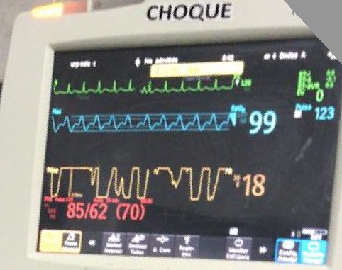
Given DKA, giving additional fluids is tempting. But before we do this, its easy to do a quick assessment of fluid tolerance #POCUS
#LUS shows some B-lines (bilat)
#IVC plethoric w no respiratory collapse
#VExUS shows very pulsatile portal vein 🚨🤔
2/12
#LUS shows some B-lines (bilat)
#IVC plethoric w no respiratory collapse
#VExUS shows very pulsatile portal vein 🚨🤔
2/12
Pulse pressure is low (23!): This suggest a low cardiac output state!
Also, there are signs of fluid intolerance!
#EchoFirst: Window is suboptimal, but we see a Hyper-dynamic LV w small cavity and a turbulent flow (green color). There was no systolic RV failure
3/12
Also, there are signs of fluid intolerance!
#EchoFirst: Window is suboptimal, but we see a Hyper-dynamic LV w small cavity and a turbulent flow (green color). There was no systolic RV failure
3/12
ICU stories: You get a call from outside 🏥 to accept a middle-aged pt w DM2/HTN/HLD/some type of solid Ca on chemo/obesity who presented to their ED w weakness/anxiety/"feeling cold". Vitals: BP 80-100, HR 130s (sinus tach), afebrile, Sat 100% on room air. Labs: WBC 13K, ...
... Lactate 5.2, creat 1.3. UA w some WBCs/bacteria. CXR clear. Norepi drip ordered but cancelled after BP improved to mid-90s, HR fell to 120s, & lactate ⬇️ to 2.5. What's your next step?
The discussion went like this:
Me: I will be happy to accept but I have no idea what we are treating. If it is sepsis, the source is unclear. And what about PE? Can you pls get a CT before sending?
ED: Sure, will do it. Thanks.
You go home & next am you learn that the CT showed:
Me: I will be happy to accept but I have no idea what we are treating. If it is sepsis, the source is unclear. And what about PE? Can you pls get a CT before sending?
ED: Sure, will do it. Thanks.
You go home & next am you learn that the CT showed:
🧵Complex #Hemodynamics in ESRD:
Middle age pt ➡️ 🏥 for syncope
HPI: Low BP during HD sessions. Does not achieve dry weight. Today she has had 6 episodes of syncope!
Last episode happened as she stood up from a chair
BP 88/62, HR 87 🧠 OK, CRT 2 sec
1/8
Middle age pt ➡️ 🏥 for syncope
HPI: Low BP during HD sessions. Does not achieve dry weight. Today she has had 6 episodes of syncope!
Last episode happened as she stood up from a chair
BP 88/62, HR 87 🧠 OK, CRT 2 sec
1/8
On #Echofist you notice this👇
During systole, flow from the LV towards the Aorta should all be blue in color! (Direction away from the probe)
This apparent change in direction (red sphere) is called Aliasing
Aliasing = Very high flow Velocity!
2/8
During systole, flow from the LV towards the Aorta should all be blue in color! (Direction away from the probe)
This apparent change in direction (red sphere) is called Aliasing
Aliasing = Very high flow Velocity!
2/8

Pt seen in ambulatory clinic with worsening kidney function
While the patient is sitting down (90 degrees), you notice neck pulsations!
Are they arterial or venous??
1/4 🧵
While the patient is sitting down (90 degrees), you notice neck pulsations!
Are they arterial or venous??
1/4 🧵
It is single peak (but not sharp)
The most striking feature is the inward movement
The breath of movement is diffuse
These are signs of venous pulsations!
Very helpful table from @AndreMansoor 👇
2/4
The most striking feature is the inward movement
The breath of movement is diffuse
These are signs of venous pulsations!
Very helpful table from @AndreMansoor 👇
2/4

Don't miss our monthly educational review #EHJACVC @ESC_Journals!
This month by the great @ArgaizR: fluids in #AKI
Co-starring: @ThinkingCC @khaycock2
Extremely proud that our journal offers a platform to 3 great clinicians & Twitter educators. I always learn from them...
This month by the great @ArgaizR: fluids in #AKI
Co-starring: @ThinkingCC @khaycock2
Extremely proud that our journal offers a platform to 3 great clinicians & Twitter educators. I always learn from them...

A strong argument is made to switch mainstream thinking in #AKI away from the fallacious concept of fluid responsiveness in all to a primary assessment of fluid tolerance.
Probably the most important thing I have learned on Twitter: #VExUS
Probably the most important thing I have learned on Twitter: #VExUS
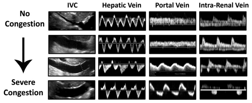
Why do I like #VExUS so much? Because it really changed my everyday practice... Portal vein became part of my standard #echocardiography assessment.
And that's what we want to achieve with this review, offer something directly applicable at your bedside!
And that's what we want to achieve with this review, offer something directly applicable at your bedside!

A short 🧵 on hepatic vein #VExUS and key pathologies
From: jacc.org/doi/10.1016/j.…
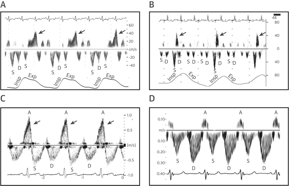
1/ HV Anatomy & Normal Flow Profile, respiratory variation (forward flow [S,D] ⬆️ during inspiration)
Click ‘ALT’ for normal waveform description
#POCUS #MedEd #Nephrology #IMPOCUS #FOAMed
From: jacc.org/doi/10.1016/j.…
1/ HV Anatomy & Normal Flow Profile, respiratory variation (forward flow [S,D] ⬆️ during inspiration)
Click ‘ALT’ for normal waveform description
#POCUS #MedEd #Nephrology #IMPOCUS #FOAMed

60 y/o ♂️ w/ ischemic heart disease & severe biventricular dysfunction. Aborted VT/VF 3x ICD shocks. Initial shock & multiorgan failure. Na 124, Cr 1.5, AST 679, ALT 765, Tbili 2.6. Una < 5mmol/L. TTE unchanged from baseline. #POCUS #vexus
What do you think is his central venous pressures based on the the IVC evaluation. Patient spontaneously breathing?
This is a very common mistake, either by tilting the transducer or getting off axis during respiratory effort. The cylinder effect is related to oblique plane insonation.
Time to update the #VExUS resources pinned thread 🧵📌
1/ Tweetorial on image acquisition.
#POCUS #MedEd
1/ Tweetorial on image acquisition.
#POCUS #MedEd
3/ Review article on #VExUS by @nephrothaniel and me in @KidneyMed
🔗kidneymedicinejournal.org/article/S2590-…
🔗kidneymedicinejournal.org/article/S2590-…

#POCUS quiz for #VExUS enthusiasts.
Image obtained from a patient with heart failure with preserved EF. IVC 1.9 cm with 30% inspiratory collapse.
Here is the intra-renal image. Interpretation of the venous waveform?
POLL in thread 👇
#MedEd #Nephrology
Image obtained from a patient with heart failure with preserved EF. IVC 1.9 cm with 30% inspiratory collapse.
Here is the intra-renal image. Interpretation of the venous waveform?
POLL in thread 👇
#MedEd #Nephrology

Young pt ➡️ 🏥 worsening shortness of breath
PMH: ESRD. Only 1 HD session/week. However, residual urine volume has now decreased substantially
On exam: BP 134/94, 2L O2,🧠✅, elevated JVP, decreased 🫁 sounds at bases, No murmurs, very mild edema. Functional left BC AVF
1/13

PMH: ESRD. Only 1 HD session/week. However, residual urine volume has now decreased substantially
On exam: BP 134/94, 2L O2,🧠✅, elevated JVP, decreased 🫁 sounds at bases, No murmurs, very mild edema. Functional left BC AVF
1/13


Careful examination of neck veins reveals no pulsations, even with pt sitting up 🤔
What could explain the absence of venous pulse? 2/13
What could explain the absence of venous pulse? 2/13
Answer is all of the above. JVP examination can be complicated in pts with ESRD.
In the absence of pulsations, I find #POCUS much helpful. Let's enhance our physical examination of congestion:
3/13


In the absence of pulsations, I find #POCUS much helpful. Let's enhance our physical examination of congestion:
3/13


#AKIConsultSeries Middle-aged male ➡️🏥 for painful knee and fever. Now in shock 🚨
📂Chart review: PMH EtOH Cirrhosis, right knee arthroplasty.
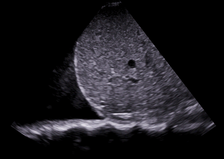
It is always a good practice review previous PACS images🩻: Nodular liver, colateral vessels and prosthetic right knee
1/11
📂Chart review: PMH EtOH Cirrhosis, right knee arthroplasty.
It is always a good practice review previous PACS images🩻: Nodular liver, colateral vessels and prosthetic right knee
1/11
On exam: BP 72/48, HR 82, O2Sat 95%.
CRT 7 sec, 🧠somnolent, confused. No edema, no obvious ascites.
Warm, swollen and erythematous knee: Tap with obvious purulent fluid🧫
Cr 2.8 mg/dl (baseline 0.5), K 6.7, Urine 🔬: hyaline casts, some urothelial cells
2/11
CRT 7 sec, 🧠somnolent, confused. No edema, no obvious ascites.
Warm, swollen and erythematous knee: Tap with obvious purulent fluid🧫
Cr 2.8 mg/dl (baseline 0.5), K 6.7, Urine 🔬: hyaline casts, some urothelial cells
2/11
Loos like hemodynamic AKI (AKA Pre-renal)
Usual causes in Cirrhosis:
🔷Distributive: Septic, "Hepatorenal physiology" 🔷Hypovolemic: Laxatives, vomiting, large volume paracentesis
🔷Congestive: Porto-pulmonary HTN, Co-existing cardiomyopathy
3/11
Usual causes in Cirrhosis:
🔷Distributive: Septic, "Hepatorenal physiology" 🔷Hypovolemic: Laxatives, vomiting, large volume paracentesis
🔷Congestive: Porto-pulmonary HTN, Co-existing cardiomyopathy
3/11
#POCUS #MedTwitter #Nephpearls
Many #VExUS enthusiasts asked for a #tweetorial on image acquisition pearls. Did one b4 but time for an updated one 🧵
#1 Let's start with basics
Color Doppler identifies the flow + tells the direction (blue is away & red towards the probe [BART])
Many #VExUS enthusiasts asked for a #tweetorial on image acquisition pearls. Did one b4 but time for an updated one 🧵
#1 Let's start with basics
Color Doppler identifies the flow + tells the direction (blue is away & red towards the probe [BART])
#2 👆BART holds good unless u invert the scale.
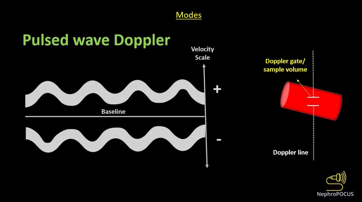
👇Pulsed wave Doppler (PWD) depicts blood flow at a certain point (sample volume) - we analyze the pattern of flow + velocity using this mode.
Above-the-baseline = flow towards the probe (like red on color)
Below = away (like blue)
👇Pulsed wave Doppler (PWD) depicts blood flow at a certain point (sample volume) - we analyze the pattern of flow + velocity using this mode.
Above-the-baseline = flow towards the probe (like red on color)
Below = away (like blue)
#3
While performing any Doppler study, it's important to keep in mind that the angle between US beam & blood flow determines the accuracy of velocity displayed. Parallel = best, perpendicular = worst
As #VExUS does not rely on absolute velocities, its OK not to have perfect angle
While performing any Doppler study, it's important to keep in mind that the angle between US beam & blood flow determines the accuracy of velocity displayed. Parallel = best, perpendicular = worst
As #VExUS does not rely on absolute velocities, its OK not to have perfect angle
#POCUS #VExUS consult for #hyponatremia. Elderly pt with h/o mitral valve replacement. On Bumetanide, UNa ~70 mmol/L, Uosm ~250🧵
Trace edema, JVD +, feels OK
Calling hemodynamic friends @khaycock2 @ThinkingCC @ArgaizR @msiuba @IM_Crit_ @MDBeni @siddharth_dugar Educate us!
1/ IVC
Trace edema, JVD +, feels OK
Calling hemodynamic friends @khaycock2 @ThinkingCC @ArgaizR @msiuba @IM_Crit_ @MDBeni @siddharth_dugar Educate us!
1/ IVC
2/ 👆Consistent with elevated right atrial pressure.
👇Hepatic vein #VExUS
D-only pattern (rhythm: ventricular paced)
👇Hepatic vein #VExUS
D-only pattern (rhythm: ventricular paced)

Se empieza a romper un poco la regla de PRIMERO LO CARGAMOS DE VOLUMEN Y DESPUÉS SI…
Al parecer va mejor iniciar un manejo en conjunto de volumen y vasopresores y así evitar la sobrecarga del paciente.
Lindo lugar para el #VeXuS
Al parecer va mejor iniciar un manejo en conjunto de volumen y vasopresores y así evitar la sobrecarga del paciente.
Lindo lugar para el #VeXuS

Small thread 🧵illustrating #POCUS based hemodynamic assessment. Relatively a classic case of pulmonary HTN and right heart failure but would like to get some insights from the experts.
1/ Parasternal long axis (PSAX) showing D-sign
#VExUS #MedEd #Nephpearls #IMPOCUS
1/ Parasternal long axis (PSAX) showing D-sign
#VExUS #MedEd #Nephpearls #IMPOCUS
2/ Parasternal long axis (PLAX) view demonstrating RV dilatation.
One of the three musketeers is big. Don't know what I'm talking about? Here is a brief reminder: 🔗nephropocus.com/2021/07/12/the…
(Mobile thing in the RVOT is PA catheter; M-mode quiz from this morning is actually this)
One of the three musketeers is big. Don't know what I'm talking about? Here is a brief reminder: 🔗nephropocus.com/2021/07/12/the…
(Mobile thing in the RVOT is PA catheter; M-mode quiz from this morning is actually this)
3/ Apical 4-chamber view #POCUS
Note how RV is dilated - bigger than LV and forming the cardiac apex.
Inter-atrial septum is bowing to the left indicating high right atrial pressure (not unexpected).
Note how RV is dilated - bigger than LV and forming the cardiac apex.
Inter-atrial septum is bowing to the left indicating high right atrial pressure (not unexpected).


