Discover and read the best of Twitter Threads about #RGphx
Most recents (10)
1/Were you today years old when you learned ASL wasn’t just short for American Sign Language?
A #tweetorial about a key perfusion method: arterial spin labeling (ASL) in collaboration w/@RadioGraphics!
Featuring this current issue article: doi.org/10.1148/rg.220…
#RGPHx
A #tweetorial about a key perfusion method: arterial spin labeling (ASL) in collaboration w/@RadioGraphics!
Featuring this current issue article: doi.org/10.1148/rg.220…
#RGPHx

2/In perfusion imaging, we want to know how blood is flowing
Usually, we do that by adding IV contrast to blood—to go along for the ride. We can track contrast by changes in MR signal
So if contrast runs w/blood, we can track blood by extension & know how it’s flowing. #RGPhx
Usually, we do that by adding IV contrast to blood—to go along for the ride. We can track contrast by changes in MR signal
So if contrast runs w/blood, we can track blood by extension & know how it’s flowing. #RGPhx

3/But what if we want to do perfusion imaging & don’t want to use contrast?
For example, in kids, we’d prefer not to give contrast.
Also, if there is an allergy, we REALLY don’t want to give contrast.
There must be another way.
#RGPhx
For example, in kids, we’d prefer not to give contrast.
Also, if there is an allergy, we REALLY don’t want to give contrast.
There must be another way.
#RGPhx

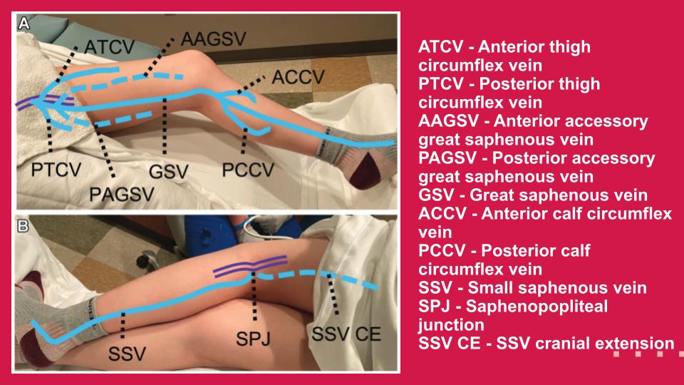
Varicose Veins are a significant cause of morbidity and healthcare expenditure. Follow along to learn more about the sonographic evaluation of venous insufficiency in the lower extremity!
@CameronAdlerMD
@MaitrayPatelMD
@cookyscan1
@RadioGraphics
@RadG_Editor
@CameronAdlerMD
@MaitrayPatelMD
@cookyscan1
@RadioGraphics
@RadG_Editor

Understanding venous anatomy is critical to performing a complete and accurate exam. Deep venous anatomy is less variable than superficial venous anatomy.
#RGphx
#RGphx

Superficial venous anatomy is more variable. Understanding the anatomy, drainage into the deep venous system, and common anatomic variants, are important.
#RGphx
#RGphx

Thread alert!
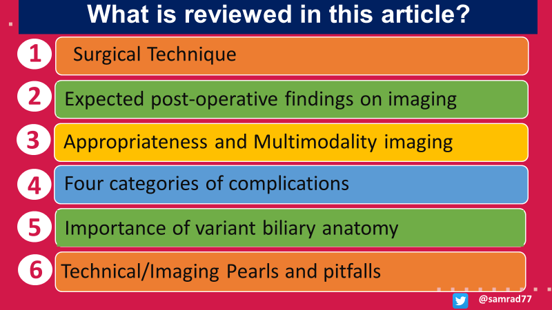
In this thread, let us learn a little more about the imaging approach to post-cholecystectomy complications.
#RGphx
@cookyscan1
#Thread
#Twittorial
1/12
In this thread, let us learn a little more about the imaging approach to post-cholecystectomy complications.
#RGphx
@cookyscan1
#Thread
#Twittorial
1/12

The latest @RadioGraphics article by @EKKpdx and her team at @OHSURadiology focuses on multimodality imaging of post-cholecystectomy complications and their management.
Link: bit.ly/3RACwQK
#RGphx @CookyScan #tweetorial
2/12
Link: bit.ly/3RACwQK
#RGphx @CookyScan #tweetorial
2/12

Cholecystectomy is one of the most performed surgeries. Radiologists and surgeons will, therefore, inevitably encounter several post-cholecystectomy complications. This article reviews role of imaging in diagnosing, preventing, and managing surgical complications.
#RGphx
3/12
#RGphx
3/12

Oy vey, the ovary! @EinsteinRads simplifies the approach to simple adnexal cysts.
Benign-appearing Incidental Adnexal Cysts at US, CT, and MR: Putting It All Together
doi.org/10.1148/rg.210…
@cookyscan1 @RadioGraphics @peterwangmd @otto07758644
#RGphx 1/9
Benign-appearing Incidental Adnexal Cysts at US, CT, and MR: Putting It All Together
doi.org/10.1148/rg.210…
@cookyscan1 @RadioGraphics @peterwangmd @otto07758644
#RGphx 1/9

Three major consensus papers were published between 2019 and 2020: the SRU Consensus Update on Adnexal Cysts, the O-RADS US Consensus Guideline, and the Management for Incidental Adnexal Findings on CT and MRI (ACR White Paper).
#RGphx 2/9
#RGphx 2/9

Most simple adnexal cysts do not require follow-up. The updated SRU guidelines provide thresholds for follow-up of 3-5 cm for postmenopausal and 5-7 cm for premenopausal patients. If a cyst decreases in size on follow-up, continued imaging is usually unnecessary.
#RGphx 3/9
#RGphx 3/9

Amyloidosis: Multisystem spectrum of Disease with Pathologic Correlation
doi.org/10.1148/rg.202…
Check out this new @RadioGraphics article by @markdsugi et al. reviewing the full spectrum of amyloidosis.
#RGphx #Tweetorial #RadRes
@markdsugi
@MSalomaoMD
@sanjeevbhalla
doi.org/10.1148/rg.202…
Check out this new @RadioGraphics article by @markdsugi et al. reviewing the full spectrum of amyloidosis.
#RGphx #Tweetorial #RadRes
@markdsugi
@MSalomaoMD
@sanjeevbhalla

AL: most common form of systemic amyloidosis in the US & seen with plasma cell dyscrasia.
AA: chronic infection/inflammation, solid organ malignancy & familial conditions.
ATTR: significant cause of morbidity and mortality related to cardiomyopathy.
#RGphx
AA: chronic infection/inflammation, solid organ malignancy & familial conditions.
ATTR: significant cause of morbidity and mortality related to cardiomyopathy.
#RGphx

CNS β-amyloid deposits ➡️cerebral amyloid angiopathy (CAA) & Alzheimer disease (AD).
CAA is an important cause of multiple intracerebral hemorrhages or cerebral microbleeds, or alternatively, a single hemorrhage in the presence of cortical superficial siderosis.
#RGphx
CAA is an important cause of multiple intracerebral hemorrhages or cerebral microbleeds, or alternatively, a single hemorrhage in the presence of cortical superficial siderosis.
#RGphx

Have you checked out the new September/October @Radiographics issue? If not, here is your #DigitalTOC with #VisualAbstracts & links for learning on the fly!
VAs designed by the talented RG VA Team.
Here we go!
#RGphx #RadRes #MedEd
@cookyscan1
1/
VAs designed by the talented RG VA Team.
Here we go!
#RGphx #RadRes #MedEd
@cookyscan1
1/
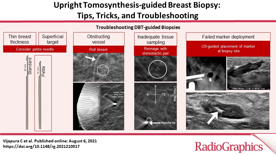
Upright Tomosynthesis-guided Breast Biopsy: Tips, Tricks, and Troubleshooting
Vijapura C et al
Breast Imaging- doi.org/10.1148/rg.202…
@CharmiMD
@RifatWahab
@MaryMahoneyMD
#RGphx
2/
Vijapura C et al
Breast Imaging- doi.org/10.1148/rg.202…
@CharmiMD
@RifatWahab
@MaryMahoneyMD
#RGphx
2/

Imaging Updates to Breast Cancer Lymph Node Management
Chung HL et al
Breast Imaging- doi.org/10.1148/rg.202…
@drhannahchung
@drjessicaleung
#RGphx
3/
Chung HL et al
Breast Imaging- doi.org/10.1148/rg.202…
@drhannahchung
@drjessicaleung
#RGphx
3/

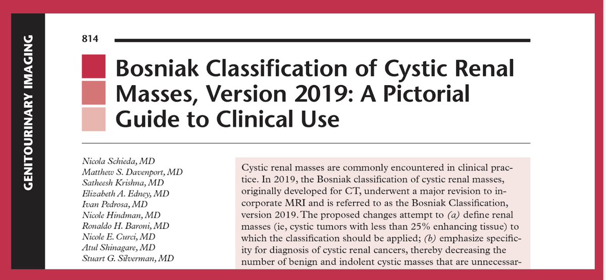
The 2019 Bosniak classification is very helpful to better characterize cystic renal masses. But there are significant changes compared to the prior edition. This pictorial by some of the brightest minds in RCC is exactly what we need to clarify some of the new changes. #RGphx 

Just to understand the reasoning for some of the changes in the Classification, let's start with a quiz; What is the estimated 10-year risk of death from a T1a (<4 cm, limited to the kidney) cystic RCC?
#RGphx @cookyscan1 @RadG_Editor
#RGphx @cookyscan1 @RadG_Editor
Given the low mortality from cystic renal masses, a greater emphasis has been put on specificity rather than sensitivity in the new edition -> a greater number of cystic renal masses are placed into lower Bosniak classes to reduce unnecessary imaging or treatment.
#RGphx
#RGphx
1 of 3 infants has a misshaped skull & 1/2500 have craniosynostosis. This new @RadioGraphics Fundamentals presentation reviews CT findings of craniosynostosis & common syndromic causes. #RGphx #radres
Craniosynostosis: Understanding the Misshaped Head bit.ly/30aRqDY
Craniosynostosis: Understanding the Misshaped Head bit.ly/30aRqDY

Before 9 months of age, only the metopic suture can be fused normally. Sagittal, coronal & lamboid sutures narrow during childhood but should not fuse. CT is imaging modality of choice- describe shape/synostosis, ICP & vascular anatomy
2/11 #RGphx
2/11 #RGphx

In non-syndromic patients, usually 1 suture is involved: sagittal > coronal > metopic > lamboid.
Let’s review the key CT features of each.
3/11 #RGphx

Let’s review the key CT features of each.
3/11 #RGphx


Traumatic pancreatic injuries are difficult to diagnose on imaging. This timely and comprehensive review article by Ayoob et al. in the latest issue of @RadioGraphics- addresses multimodality imaging of pancreatic trauma.
1/19
#RGphx
#RadRes #RadEd #tweetorial
@CookyScan
1/19
#RGphx
#RadRes #RadEd #tweetorial
@CookyScan

Pancreatic injuries are frequently overlooked on initial CT due to the low frequency of these injuries, associated nonspecific clinical features, and relatively subtle imaging findings, and the presence of associated multiple organ injuries.
2/19 #RGphx
2/19 #RGphx
In a patient with abdominal organ injuries on CT, how frequently is a pancreatic injury associated?
3/19 #RGphx
3/19 #RGphx
DECT- An innovative CT technology, produces numerous imaging datasets at >1 energy level. The image reconstruction algorithms lead to pitfalls, the radiologists must be aware of. A #Tweetorial thread
@RadioGraphics @cookyscan1
#RGphx
doi.org/10.1148/rg.202…
@RadioGraphics @cookyscan1
#RGphx
doi.org/10.1148/rg.202…

DECT enables the synthesis of a wide array of images from a single acquisition because of the availability of CT projection data at more than one polychromatic energy level. Currently, six major approaches are used for DECT.
@RadioGraphics @cookyscan1
#RGphx

@RadioGraphics @cookyscan1
#RGphx


VMC images-specific to DECT, available with all platforms at multiple keV levels starting from 40 keV. Images at 40–70 keV remain susceptible to pseudo enhancement similar to that on polychromatic single-energy CT images @RadioGraphics @cookyscan1
#RGphx

#RGphx


